Posts tagged ultrasound
Intern Ultrasound of the Month: Posterior Vitreous Detachment

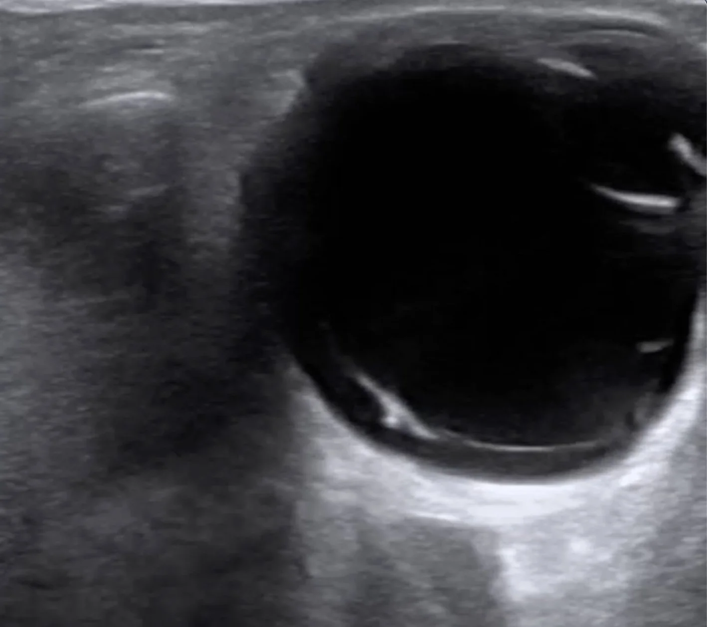
This post by Dr. Marco Lorico Rappa highlights the classic POCUS appearance of posterior vitreous detachment, reviews key sonographic features that distinguish it from retinal detachment and vitreous hemorrhage, and discusses practical scanning techniques — including gain optimization and dynamic oculokinetic imaging — to improve diagnostic accuracy.

Read More
Intern Ultrasound of the Month: Detection of Aortic Dissection During Cardiac Arrest

This post by Dr. Joanna Dehghani highlights the role of POCUS during cardiac arrest, demonstrating how a systematic ultrasound evaluation can uncover catastrophic pathology beyond the usual reversible causes. Learn how bedside ultrasound can aid in the diagnosis of acute aortic dissection, recognize key sonographic findings, and guide critical management decisions.

Read More
Intern Ultrasound of the Month: Lipomatous Atrial Septal Hypertrophy (LASH) & Other Cardiac Masses

Lipomatous atrial septal hypertrophy (LASH) is an uncommon, benign finding that can easily be mistaken for more serious cardiac pathology. This post by Dr. Ashley Timmreck-Walker reviews the characteristic POCUS features of LASH and highlights key imaging pearls to help differentiate it from other intracardiac masses.

Read More
Ultrasound-Guided Pericardiocentesis: A Review

This post by Dr. Thomas Evans walks through the essentials of ultrasound-guided pericardiocentesis — when to do it, how to do it, and what pitfalls to avoid. We highlight the subxiphoid, parasternal, and apical approaches with practical POCUS tips for real-time needle visualization, plus a review of complications and how to keep the procedure as safe as possible.

Read More
Intern Ultrasound of the Month: Cardiac Tamponade

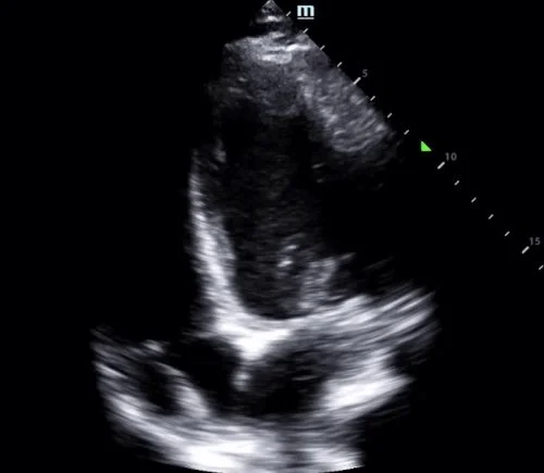
This case is by Dr. Ryan Starkman (now PGY2) and is a great example of a large circumferential pericardial effusion with sonographic tamponade physiology diagnosed with POCUS. This expedited interventional cardiology involvement before the patient decompensated, and 750cc of fluid was successfully drained in the cath lab.

Read More
Intern Ultrasound of the Month: Retinal Detachment

This month’s case is by Dr. Yanina Guevara who shares a case of painless monocular vision changes in a patient who was found to have a retinal detachment (and vitreous hemorrhage), first diagnosed with point-of-care ultrasound! This is accompanied by an overview of how to perform an ultrasound ocular exam and highlights some of the pathologies that can be visualized. Read on to learn more!

Read More