Posts tagged vitreous detachment
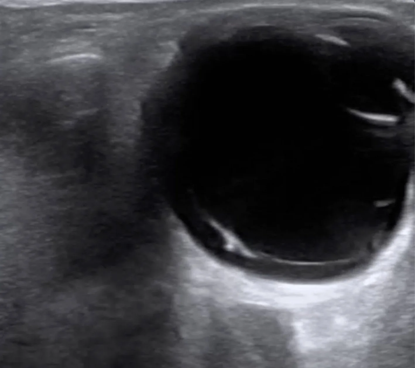
Intern Ultrasound of the Month: Posterior Vitreous Detachment

This post by Dr. Marco Lorico Rappa highlights the classic POCUS appearance of posterior vitreous detachment, reviews key sonographic features that distinguish it from retinal detachment and vitreous hemorrhage, and discusses practical scanning techniques — including gain optimization and dynamic oculokinetic imaging — to improve diagnostic accuracy.

Read More
Intern Ultrasound of the Month: Retinal Detachment

This month’s case is by Dr. Yanina Guevara who shares a case of painless monocular vision changes in a patient who was found to have a retinal detachment (and vitreous hemorrhage), first diagnosed with point-of-care ultrasound! This is accompanied by an overview of how to perform an ultrasound ocular exam and highlights some of the pathologies that can be visualized. Read on to learn more!

Read More