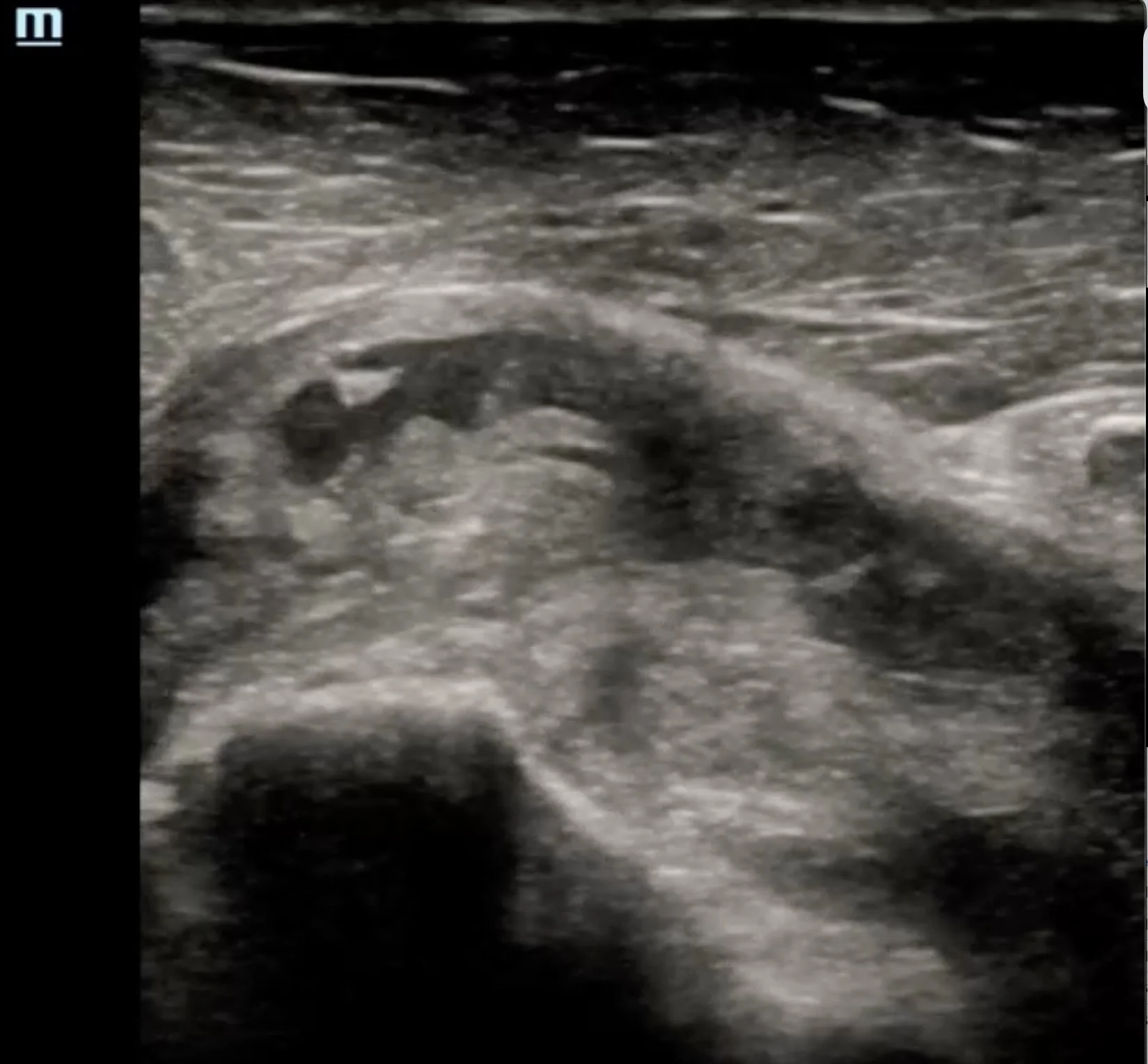
This blog reviews the point-of-care ultrasound evaluation of the rotator cuff, including patient positioning, probe placement, image acquisition, normal sonoanatomy, and common pathology. By the end, you'll have a systematic approach to performing a comprehensive rotator cuff examination at the bedside.
Read MoreDeep vein thrombosis (DVT) is a common and potentially life-threatening condition. In this post by Dr. Suela Lamaj, we review the indications, image acquisition, interpretation, and key pearls for performing lower extremity DVT POCUS in the emergency department.
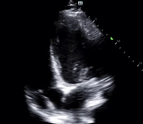
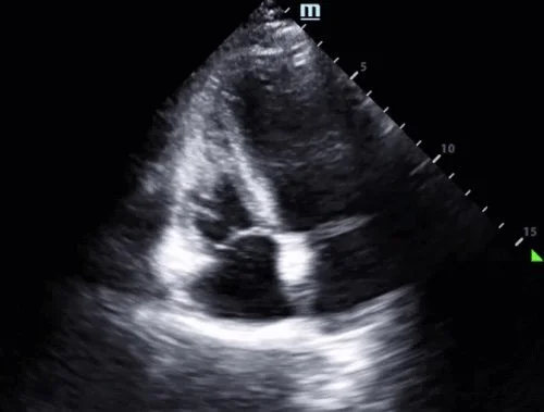
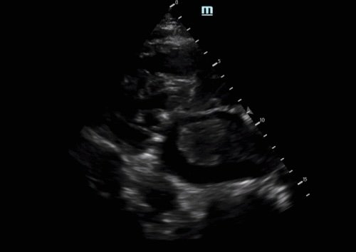
Read MoreThis blog post by Dr. Kyle Copp highlights a case of ICD lead dislodgment identified with POCUS. Learn how a focused cardiac ultrasound exam, including the apical four-chamber and modified parasternal views, can rapidly identify lead malposition, complement device interrogation, and expedite definitive management.
Read MoreThis post by Dr. Marco Lorico Rappa highlights the classic POCUS appearance of posterior vitreous detachment, reviews key sonographic features that distinguish it from retinal detachment and vitreous hemorrhage, and discusses practical scanning techniques — including gain optimization and dynamic oculokinetic imaging — to improve diagnostic accuracy.
Read MoreThis post by Dr. Joanna Dehghani highlights the role of POCUS during cardiac arrest, demonstrating how a systematic ultrasound evaluation can uncover catastrophic pathology beyond the usual reversible causes. Learn how bedside ultrasound can aid in the diagnosis of acute aortic dissection, recognize key sonographic findings, and guide critical management decisions.
Read MoreIn this post by Dr. Anna Wobser, we review a trauma case of placental abruption and discuss the sonographic findings, image acquisition, and common pitfalls to help clinicians identify this high-risk, time-sensitive obstetric emergency at the bedside.
Read MoreLipomatous atrial septal hypertrophy (LASH) is an uncommon, benign finding that can easily be mistaken for more serious cardiac pathology. This post by Dr. Ashley Timmreck-Walker reviews the characteristic POCUS features of LASH and highlights key imaging pearls to help differentiate it from other intracardiac masses.
Read MoreLeft atrial thrombus is a rare but important POCUS finding with significant implications for patient care. This post by Dr. Ryan Schroeder reviews the key ultrasound features, differential diagnosis, and clinical considerations to help you recognize this potentially life-threatening pathology.
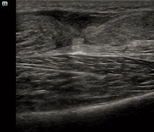
Read MoreThis blog post by Dr. Calvin Jackson highlights a case of Achilles tendon rupture, a common cause of acute posterior ankle pain in the emergency department. Learn how POCUS can rapidly confirm the diagnosis, recognize key sonographic findings, avoid common pitfalls, and facilitate timely orthopedic consultation.
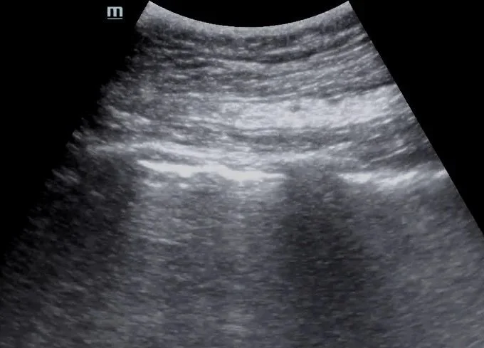
Read MoreThis post by Dr. Jordan DeAngelis reviews the use of point-of-care lung ultrasound to assess for pneumothorax, focusing on image acquisition, interpretation of lung sliding and the lung point, and practical tips to avoid common pitfalls.
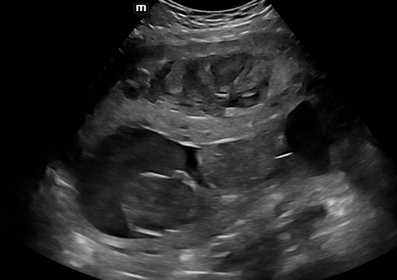
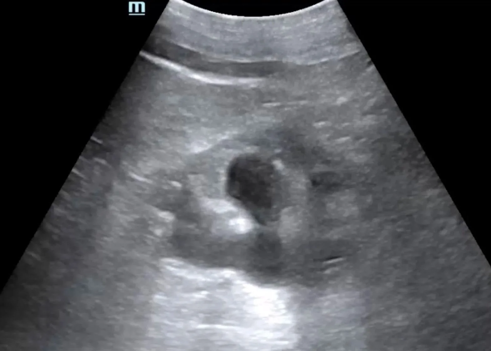
Read MoreThis case is by Dr. Nicole Minnerath and illustrates the detection of new hydronephrosis in a patient with bilateral nephrostomy tubes and ureteral stents. Read on to explore the role of renal and bladder POCUS in evaluating obstructive uropathy.
Read MoreIn this post, Dr. Tiana Sarsour highlights a case of recurrent abscesses and fever where POCUS confirmed a drainable collection, followed by a great review of abscess management and the role of ultrasound for skin and soft tissue infections.
Read MoreThis case is by Dr. Hannah Hendrix (now PGY2) and features a great example of the retrobulbar spot sign — a unique finding associated with embolic central retinal artery occlusion (CRAO). Read on for a quick review of ocular ultrasound and a closer look at this highly specific sonographic feature.
Read MoreThis post by Dr. Thomas Evans walks through the essentials of ultrasound-guided pericardiocentesis — when to do it, how to do it, and what pitfalls to avoid. We highlight the subxiphoid, parasternal, and apical approaches with practical POCUS tips for real-time needle visualization, plus a review of complications and how to keep the procedure as safe as possible.
Read MoreThis case is by Dr. Ryan Starkman (now PGY2) and is a great example of a large circumferential pericardial effusion with sonographic tamponade physiology diagnosed with POCUS. This expedited interventional cardiology involvement before the patient decompensated, and 750cc of fluid was successfully drained in the cath lab.
Read MoreThis case is by Dr. Jacob Perino and is a great example of an incidental finding of an iliac aneurysm found when performing a bladder ultrasound on a patient presenting with a malfunctioning Foley catheter. Read on to learn more about iliac aneurysms and bladder ultrasound in the setting of a Foley.
Read MoreThis month’s case is by Dr. Yanina Guevara who shares a case of painless monocular vision changes in a patient who was found to have a retinal detachment (and vitreous hemorrhage), first diagnosed with point-of-care ultrasound! This is accompanied by an overview of how to perform an ultrasound ocular exam and highlights some of the pathologies that can be visualized. Read on to learn more!
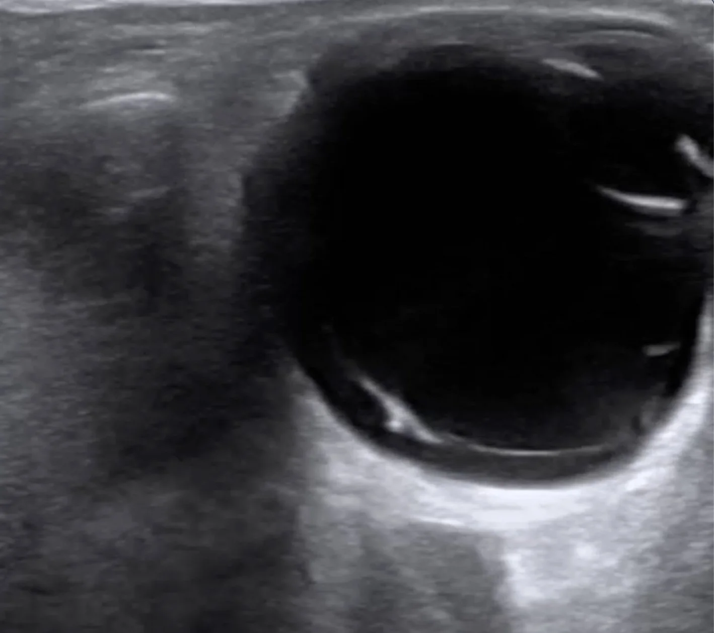
Read MoreOur latest Intern Ultrasound of the Month is by Dr. Abby Wissman and features a cool case of a complex cystic renal mass found incidentally. She reviews the basics of renal POCUS and dives into a great discussion about various renal pathology that we may see on ultrasound.
Read MoreTo follow up our most recent case of McConnell’s sign in the setting of an acute pulmonary embolism, Dr. Isma Dhanani goes more in-depth on the right heart and covers additional signs of right heart dysfunction - acute and chronic - that can be quickly identified with POCUS.
Read MoreOur latest Intern Ultrasound of the Month is brought to you by Dr. Gabe Alagna. He presents a case in which POCUS performed on a patient in cardiac arrest found an enlarged right heart with McConnell’s sign, raising concern for PE, which quickly changed management. Read on to learn more!
Read More