Intern Ultrasound of the Month: Don't Overlook the Right Ventricle!
The Case
55-year-old male with a past medical history of hypertension and diabetes presented to the emergency department for shortness of breath and dizziness. He reported these symptoms started while he was exerting himself and minimally improved after a period of rest. He reported feeling like he was going to pass out but did not experience loss of consciousness. The patient denied any chest pain, headache, fever, chills, recent illnesses, or nausea/vomiting. He reported similar symptoms in the past without further workup or diagnosis.
His vitals were within normal limits and his examination was unremarkable.
In evaluating his pre-syncopal episode, an EKG was obtained and showed sinus rhythm at a rate of 70 with right axis deviation and T-wave inversions in anteroseptal leads without acute ST elevations, similar to a previous EKG from several months ago.
While awaiting a chest x-ray and labs, a cardiac point-of-care ultrasound (POCUS) was performed.

Parasternal long axis view - The RV appears dilated and is relatively larger than the aortic outflow tract and left atrium; normally these should have ~1:1:1 ratio.

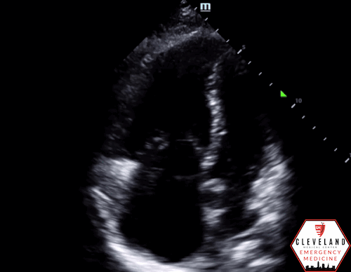
Parasternal short axis view - The RV appears larger than the LV and there is flattening/bowing of the interventricular septum (“D sign”).
Apical four chamber view - The right-sided cardiac chambers (screen left) are notably larger than the left-sided chambers (screen right).

Tricuspid annular plane systolic excursion (TAPSE) - a quantitative assessment of vertical movement of the tricuspid annulus, which reflects RV function (see below to learn more). Here, the TAPSE is less than 17mm, indicating reduced RV function.

Inferior vena cava (IVC) - The IVC is plethoric with reduced respiratory variation, an adjunct to cardiac findings that further supports evidence of right heart dysfunction.
Overall impression of POCUS findings:
Right heart enlargement with septal flattening/bowing (“D sign”) in the parasternal short axis view and reduced TAPSE, indicating reduced RV function. The IVC is also plethoric, which further supports this. LV function is preserved and there’s no pericardial effusion.
Case Continued: Labs were significant for a slightly elevated BNP of 300. COVID PCR was negative. Chest x-ray showed mild pulmonary vascular congestion. On review of prior records, the patient had previously been diagnosed with pulmonary hypertension, and a recent echo had similar findings. Given the patient’s overall stability, the ED team held off on additional imaging and admitted him for further evaluation and management.
Right Heart Dysfunction/Failure
Brief Background/Overview
Often overshadowed by the left ventricle (LV), the right ventricle (RV) is a unique chamber of the heart with distinct anatomical and physiologic features. It has a complex shape with longitudinally-oriented muscle fibers whose shortening contributes to much of its contractility (1). Compared to the LV, the RV is thinner and composed of fewer muscle fibers. This is because the pressure in the pulmonary circuit is much lower than that of systemic circulation, allowing the RV to maintain the same stroke volume as the LV but with 75% less stroke work (2). The RV is also more compliant than the LV, which allows the RV to accommodate the large volume fluctuations of venous return without affecting the end-diastolic pressure (2). In contrast, the RV does not withstand an acute pressure load well and is highly sensitive to pressure changes (1). Despite their differences, the RV and LV share the interventricular septum as well as the pericardial space. As a result, they are interdependent, and dysfunction of one often leads to dysfunction of the other (3).
RV function is largely influenced by preload, afterload, and contractility [1]. Dysfunction is defined as abnormal structure or function of the RV, while right heart failure (RHF) refers to a clinical syndrome that can develop from RV dysfunction (4). Potential causes include pulmonary arterial hypertension, massive pulmonary embolism, right-sided myocardial infarction, acute respiratory distress syndrome (ARDS), chronic obstructive pulmonary disease (COPD), chronic lung disease, RV outflow obstruction, myocarditis, and advanced LV failure, to name a few. These pathologies lead to RV dysfunction/failure through pressure overload, volume overload, or decreased contractility of the right heart (1,3,4).
Because RV function is highly sensitive to changes in afterload, even slight increases - especially sudden onset, are poorly tolerated and result in significant changes in stroke volume. Gradual increases in RV afterload may allow the ventricle time to adapt, though it may still eventually lead to RV dysfunction (5). Reduced RV stroke volume leads to RV dilation, which causes tricuspid regurgitation, further exacerbating RV dilation. Due to ventricular interdependence, this leads to impaired LV filling and reduced cardiac output (4).
Clinical Presentation
The clinical presentation of right heart failure largely depends on the underlying cause and severity of the RV dysfunction. Some of the common signs and symptoms include shortness of breath with exertion, peripheral edema, fatigue, jugular venous distention, hepatomegaly, hepatojugular reflux, and ascites (1,3). In advanced cases of RV dysfunction, patients can present with hypotension or shock and signs of hypoperfusion (4).
Diagnosis
The diagnosis of RV dysfunction depends on the patient’s clinical presentation, examination findings, EKG findings, and imaging findings. On EKG, there may be evidence of right axis deviation and right ventricular hypertrophy. If the cause of RV dysfunction is due to a massive PE, there may be the classically-associated “S1Q3T3”, or McGinn-White Sign, due to acute cor pulmonale; this sign is comprised of an S wave in lead I, a Q wave in lead III, and an inverted T wave in lead III (2). X-rays, which are commonly ordered given the clinical presentation, may show signs such as cardiomegaly or pulmonary edema, though findings are not specific to RV dysfunction (4).
Most commonly, RV dysfunction is diagnosed by comprehensive echocardiography. It is a non-invasive study that can assess the size, morphology, and function of the right heart, pulmonary hemodynamics, and associated structures (4,6). While not comprehensive, POCUS allows for a rapid assessment of RV size, global function, the presence of tricuspid regurgitation or pericardial effusion, and inferior vena cava (IVC) size and collapsibility (6).
Cardiac MRI is the gold standard for evaluating for RV abnormalities because it allows for good visualization of anatomy, tissue characterization, calculation of flow, and quantification of function (3). However, MRIs are costly, not always readily available, and may also cause malfunction of implanted devices (such as pacemakers), thus resulting in echocardiography being the initial diagnostic modality of choice (6).
Management
Management of RV dysfunction/failure largely depends on the underlying etiology. Treating the underlying cause and triggering factors is key. The overarching goals of treatment include optimizing RV preload, reducing RV afterload, and increasing RV contractility and perfusion (4,5,7). To optimize preload, assess and carefully support volume status as needed. In the setting of fluid overload, diuretics can be used to decrease venous congestion as well as relieve hypoxia. To reduce RV afterload, first aim to correct hypoxia, acidemia, and hypercapnia, as these can increase pulmonary vascular resistance. In advanced cases, afterload-reducing agents, i.e. prostacycline and other pulmonary vasodilators, may be indicated. Vasopressors can be used to increase systemic arterial blood pressure and improve perfusion. Inotropes, such as Dobutamine or Milrinone, can improve myocardial contractility while also improving afterload; however, these can also reduce systemic vascular resistance so systemic vasoconstrictors may also be needed to counteract this (5,7).
Complications of right heart failure, especially if untreated, include end-organ damage. Primarily affecting the liver and kidneys, this results from increased venous pressure and reduced perfusion from the decreased cardiac output (4).
Evaluating the Right Heart with POCUS
In patients with shortness of breath, hypotension, syncope/near-syncope, JVD, etc, a focused evaluation of the heart with POCUS can provide a lot of information at bedside, which can be especially useful in unstable patients. Specifically when concerned about right heart pathology, POCUS allows us to quickly assess RV size, global function, the presence of a pericardial effusion, gross valvular abnormalities and regurgitation, and IVC size and collapsibility (8,9). Findings are predominantly described qualitatively (normal, mild, severe) or with a yes/no answer, which the clinician can correlate with and apply to the clinical picture. It is important to note that POCUS does have its limitations and should not replace a comprehensive study.
RV Enlargement (RV: LV ratio)
For POCUS, the most basic component to evaluate is RV size, especially relative to the LV. This can be assessed qualitatively in an apical four chamber view. The RV should be smaller than the LV, so if the RV is similar in size or larger than the LV, this indicates enlargement (10). POCUS performed by emergency physicians can accurately identify RV dysfunction with high sensitivity and specificity, outperforming cardiac biomarkers and CT findings (11).

Figure 1. RV: LV ratio in an apical four chamber view. If the ratio is >1, this indicates RV enlargement.

Figure 2. “D sign” in a parasternal short axis view, indicating pressure or volume overload on the RV.

Figure 3. Eccentricity Index. Source: POCUS 101.
“D Sign”
Seen on a parasternal short axis view, this is characterized by flattening/leftward deviation of the interventricular septum so that the LV has a “D” shape, see Figure 2. This is a result of overload on the RV (10). To determine whether it is pressure or volume overload is causing this RV strain, you can calculate the eccentricity index (EI).
—> Eccentricity Index
Described by Ryan et al., this consists of measuring the LV diameter parallel and perpendicular to the septum in a parasternal short axis view. Do this in both end-systole and end-diastole and then calculate the ratio for each. See Figure 3.
EI = D2/D1 (D2 = LV diameter parallel to the septum, D1 = LV diameter perpendicular to the septum in parasternal short-axis.
Normal EI ≤ 1 (the LV is circular)
RV volume overload: EI > 1 during end-diastole but EI ≤ 1 (normal) during end-systole
RV pressure overload: EI >1 during BOTH end-systole & end-diastole (12).

Figure 4. TAPSE, indicating reduced RV function
Tricuspid Annular Plane Systolic Excursion (TAPSE)
Because the majority of the RV muscle fibers contract longitudinally, the vertical motion of the RV roughly indicates RV function (3). The greater the vertical displacement of the tricuspid annulus during systole, the better the function. This can be assessed in an apical four chamber view. Using m-mode, place the bar over the lateral tricuspid annulus. On the waveform produced, measure the distance from the peak to trough, see Figure 4. A measurement less than 17 mm is considered abnormal, indicating reduced RV function. It is important to note that this is angle-dependent so if the view is off-axis or if the m-mode bar is not well-aligned, it can result in an erroneous measurement (13).

Figure 5. RV wall thickness; >5mm indicates a chronic process.
RV Wall Thickness
If the thickness of the RV free wall is greater than 5mm, it indicates RV hypertrophy and suggests a chronic process (though it does not exclude a concurrent acute one). This is best measured in parasternal long axis view or subxiphoid view at end-diastole, see Figure 5 (13).
IVC
A quick assessment of the IVC is another adjunct to aforementioned views. A plethoric, or distended, IVC with minimal respiratory variation within roughly 3 cm of the right atrium has been shown to reflect increased right atrial pressure (10).
While not covered in this blog post, there are several additional signs of RV strain that can be evaluated with POCUS. This includes McConnell’s sign (see previous blog post for details) and other quantitative measurements of right ventricular systolic pressure, pulmonary artery pressure and acceleration time, tricuspid regurgitation, among others. See additional resources below to learn more.
Take Home Points
A patient with right heart failure can present with non-specific symptoms but POCUS can quickly, non-invasively, and effectively help identify and narrow the differential.
POCUS can identify signs of RV dysfunction, including RV enlargement, a “D” sign, reduced TAPSE.
To differentiate volume vs pressure overload causing a “D" sign”, calculate the eccentricity index — septal flattening in diastole occurs in both but only in pressure overload states does this also occur in systole.
The mainstay of treatment is focused on optimizing preload, reducing afterload, and improving contractility and perfusion while addressing the underlying cause.
Early recognition of RV dysfunction can help initiate and guide appropriate management, monitor progression, and improve morbidity and mortality.
AUTHORED BY: ISMA DHANANI, MD, PGY1
FACULTY CO-AUTHOR/EDITOR: LAUREN MCCAFFERTY, MD
References
Dini FL, Pugliese NR, Ameri P, et al. Heart Failure Study Group of the Italian Society of Cardiology. Right ventricular failure in left heart disease: from pathophysiology to clinical manifestations and prognosis. Heart Fail Rev. 2023; 28(4):757-766.
Arrigo M, Huber LC, Winnik S, et al. Right Ventricular Failure: Pathophysiology, Diagnosis and Treatment. Card Fail Rev. 2019;5(3):140-146
Ibrahim BS. Right ventricular failure. ESC Escardio.org. 2016; 14(32). https://www.escardio.org/Journals/E-Journal-of-Cardiology-Practice/Volume-14/Right-ventricular-failure.
Konstam MA, Kiernan MS, Bernstein D, et al. Evaluation and Management of Right-Sided Heart Failure: A Scientific Statement From the American Heart Association. Circulation. 2018;137(20):e578-e622.
Murphy E, Shelley B. Clinical presentation and management of right ventricular dysfunction. BJA Educ. 2019;19(6):183-190.
Ro SK, Sato K, Ijuin S, et al. Assessment and diagnosis of right ventricular failure-retrospection and future directions. Front Cardiovasc Med. 2023;10:1030864.
Ventetuolo CE, Klinger JR. Management of acute right ventricular failure in the intensive care unit. Ann Am Thorac Soc. 2014;11(5):811-22.
Johri AM, Galen B, Kirkpatrick JN, Lanspa M, Mulvagh S, Thamman R. ASE Statement on Point-of-Care Ultrasound during the 2019 Novel Coronavirus Pandemic. J Am Soc Echocardiogr. 2020; 33(6):670-673.
Spencer KT, Flachskampf FA. Focused Cardiac Ultrasonography. JACC Cardiovasc Imaging. 2019; 12(7 Pt 1):1243-1253.
Reardon RF, Laudenbach A, Joing SA. Cardiac. In OJ Ma, JR Mateer, RF Reardon, SA Joing (eds), Ma and Mateer’s Emergency Ultrasound (3rd ed). 2008. New York, NY: McGraw-Hill Education. pp 93-167.
Weekes AJ, Thacker G, Troha D, Johnson AK, et al. Diagnostic Accuracy of Right Ventricular Dysfunction Markers in Normotensive Emergency Department Patients With Acute Pulmonary Embolism. Ann Emerg Med. 2016;68(3):277-91.
Ryan T, Petrovic O, Dillon JC, Feigenbaum H, Conley MJ, Armstrong WF. An echocardiographic index for separation of right ventricular volume and pressure overload. J Am Coll Cardiol. 1985; 5(4):918-27.
Rudski LG, Lai WW, Afilalo J, Hua L, Handschumacher MD, Chandrasekaran K, Solomon SD, Louie EK, Schiller NB. Guidelines for the echocardiographic assessment of the right heart in adults: a report from the American Society of Echocardiography endorsed by the European Association of Echocardiography, a registered branch of the European Society of Cardiology, and the Canadian Society of Echocardiography. J Am Soc Echocardiogr. 2010;23(7):685-713.
To learn more, check out these additional resources:
https://www.pocus101.com/the-d-sign-right-heart-strain-from-pressure-vs-volume-overload/
https://coreultrasound.com/right-heart-function/
https://coreultrasound.com/60-60-sign-for-acute-pulmonary-embolism/