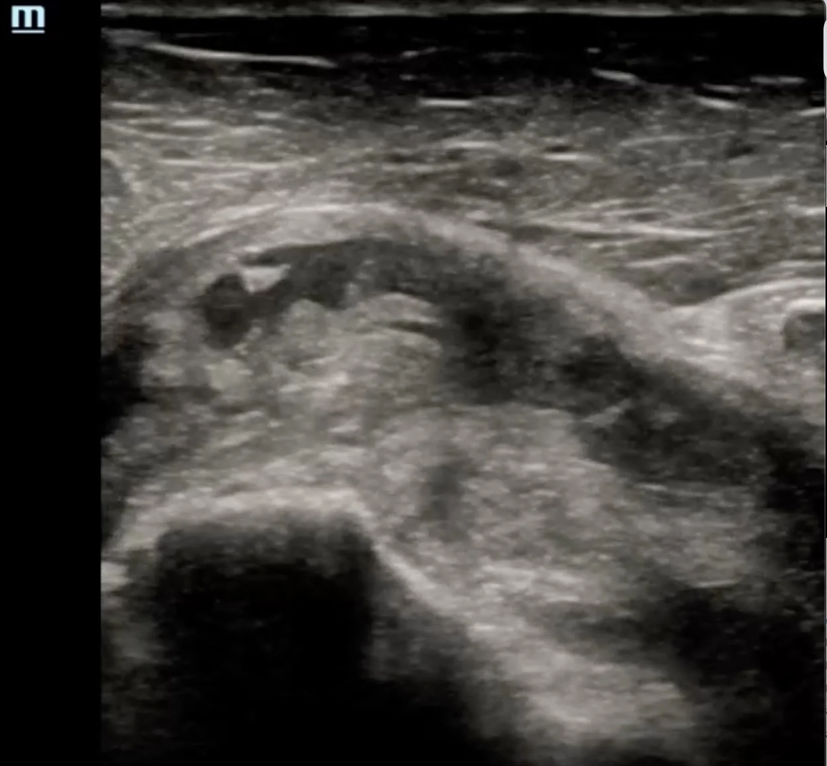
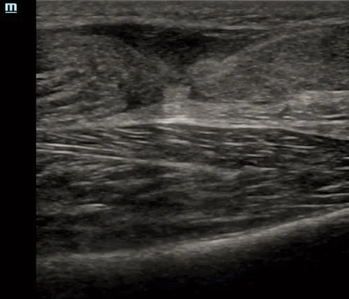
This blog reviews the point-of-care ultrasound evaluation of the rotator cuff, including patient positioning, probe placement, image acquisition, normal sonoanatomy, and common pathology. By the end, you'll have a systematic approach to performing a comprehensive rotator cuff examination at the bedside.
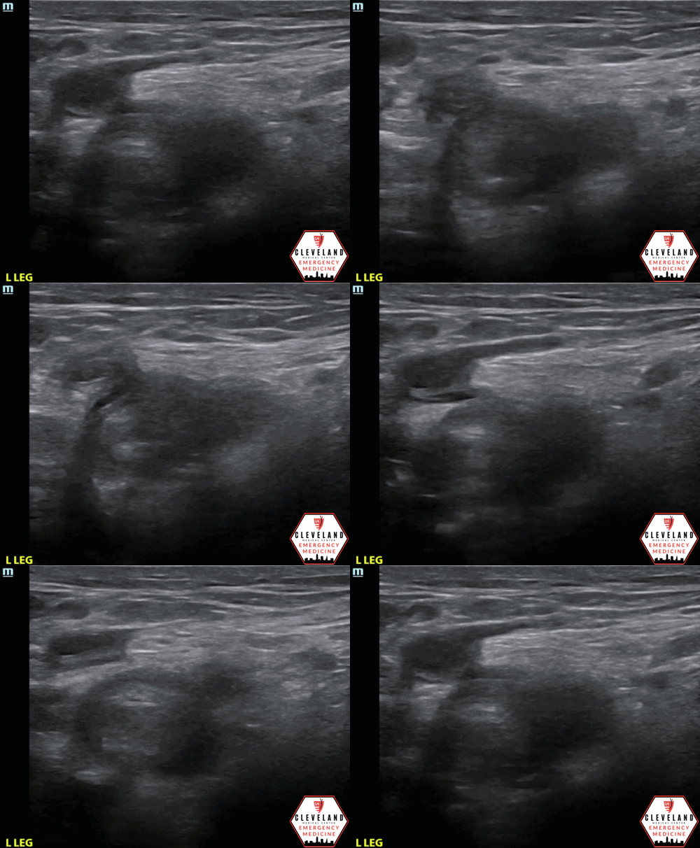
Deep vein thrombosis (DVT) is a common and potentially life-threatening condition. In this post by Dr. Suela Lamaj, we review the indications, image acquisition, interpretation, and key pearls for performing lower extremity DVT POCUS in the emergency department.
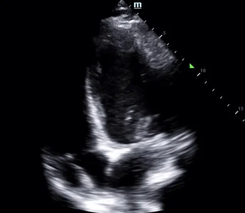
This blog post by Dr. Kyle Copp highlights a case of ICD lead dislodgment identified with POCUS. Learn how a focused cardiac ultrasound exam, including the apical four-chamber and modified parasternal views, can rapidly identify lead malposition, complement device interrogation, and expedite definitive management.
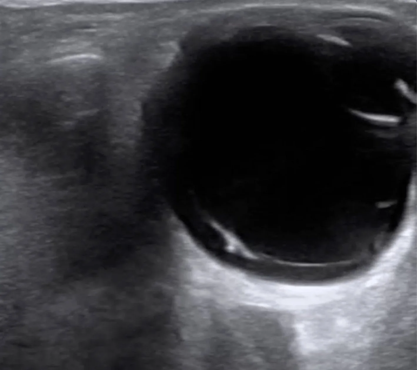
This post by Dr. Marco Lorico Rappa highlights the classic POCUS appearance of posterior vitreous detachment, reviews key sonographic features that distinguish it from retinal detachment and vitreous hemorrhage, and discusses practical scanning techniques — including gain optimization and dynamic oculokinetic imaging — to improve diagnostic accuracy.
This post by Dr. Joanna Dehghani highlights the role of POCUS during cardiac arrest, demonstrating how a systematic ultrasound evaluation can uncover catastrophic pathology beyond the usual reversible causes. Learn how bedside ultrasound can aid in the diagnosis of acute aortic dissection, recognize key sonographic findings, and guide critical management decisions.
In this post by Dr. Anna Wobser, we review a trauma case of placental abruption and discuss the sonographic findings, image acquisition, and common pitfalls to help clinicians identify this high-risk, time-sensitive obstetric emergency at the bedside.
This blog post from medical student Kenna Hawes, MS4 CWRU SOM, provides a great review of cyanobacterial harmful algal blooms. Learn more about how cyanobacterial toxins affect the body, recognize the clinical manifestations of exposure, and apply key diagnostic and management principles for this increasingly important environmental exposure.
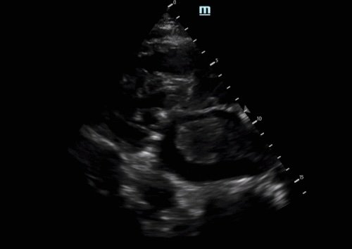
Lipomatous atrial septal hypertrophy (LASH) is an uncommon, benign finding that can easily be mistaken for more serious cardiac pathology. This post by Dr. Ashley Timmreck-Walker reviews the characteristic POCUS features of LASH and highlights key imaging pearls to help differentiate it from other intracardiac masses.
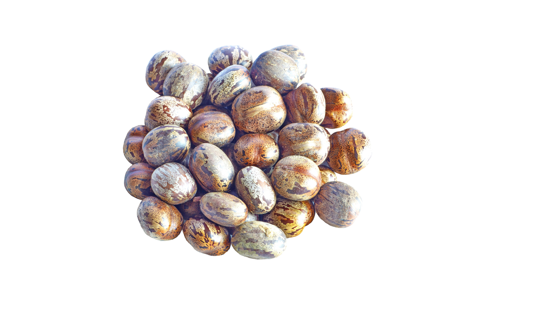
This blog post from medical student Lisa Tan is an excellent review of ricin toxicity. Learn more about how this potent toxin presents, why the route of exposure matters, and the key management pearls every emergency clinician should know.
Left atrial thrombus is a rare but important POCUS finding with significant implications for patient care. This post by Dr. Ryan Schroeder reviews the key ultrasound features, differential diagnosis, and clinical considerations to help you recognize this potentially life-threatening pathology.
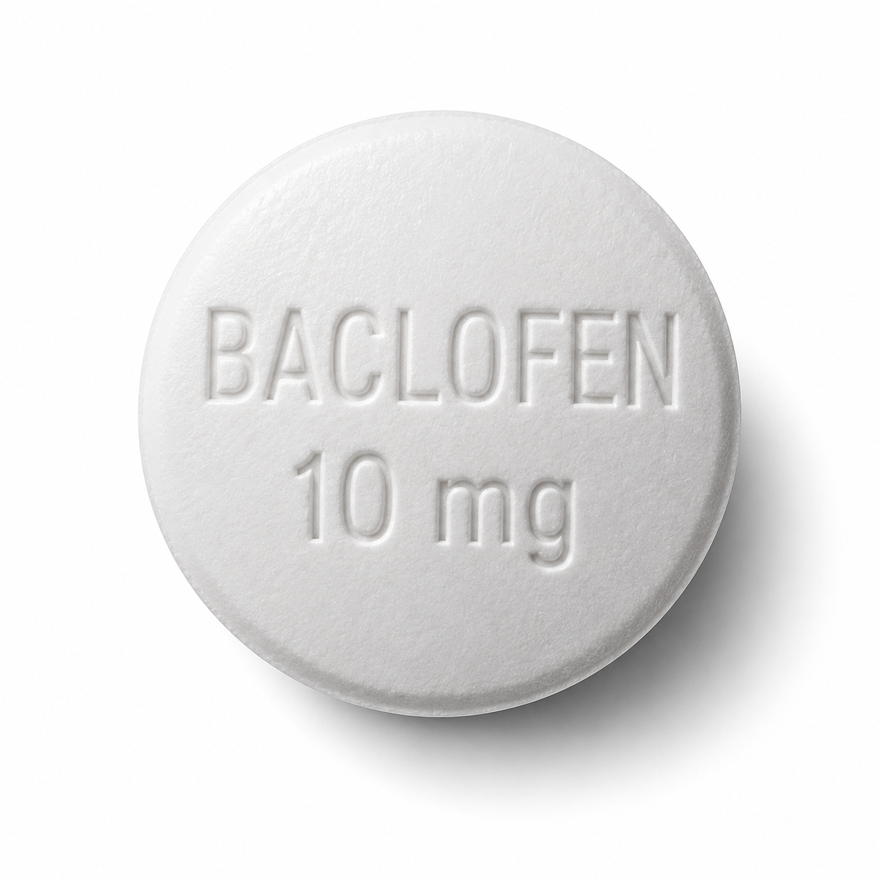
This blog post from last year's residency grad, Dr. Kelsie Rhyne, is an excellent review of baclofen toxicity. Learn more about how to recognize this potentially life-threatening toxidrome, navigate diagnostic challenges, and apply key management pearls.
This blog post is from last year’s grad, Dr. Vince Marshall — a concise review of vinyl chloride toxicity, emphasizing exposure risks, acute and chronic effects, and emergency care.
Check out our latest tox blog post by current chief, Dr. Jake Perino, highlighting the history, pathophysiology, and key management pearls of salicylate toxicity.
This blog post by Dr. Calvin Jackson highlights a case of Achilles tendon rupture, a common cause of acute posterior ankle pain in the emergency department. Learn how POCUS can rapidly confirm the diagnosis, recognize key sonographic findings, avoid common pitfalls, and facilitate timely orthopedic consultation.
Kratom use has been increasing and political conversations regarding its legality are ever-present in the daily news cycle. Our chief resident Dr. Wissman will discuss Kratom and its presentation and toxicity. Enjoy and remember to call your poison center!
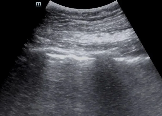
This post by Dr. Jordan DeAngelis reviews the use of point-of-care lung ultrasound to assess for pneumothorax, focusing on image acquisition, interpretation of lung sliding and the lung point, and practical tips to avoid common pitfalls.
Check out this blog post by Grace Van Hare, MS4, on three high-yield uses of agitated saline with POCUS in resuscitation. From central line confirmation and RV dysfunction to shunt detection, it offers a practical, bedside-focused discussion.
Check out our latest Resus blog post! Megan Callaghan, MS4, breaks down Sympathetic Crashing Acute Pulmonary Edema (SCAPE) — from clinical presentation to management.
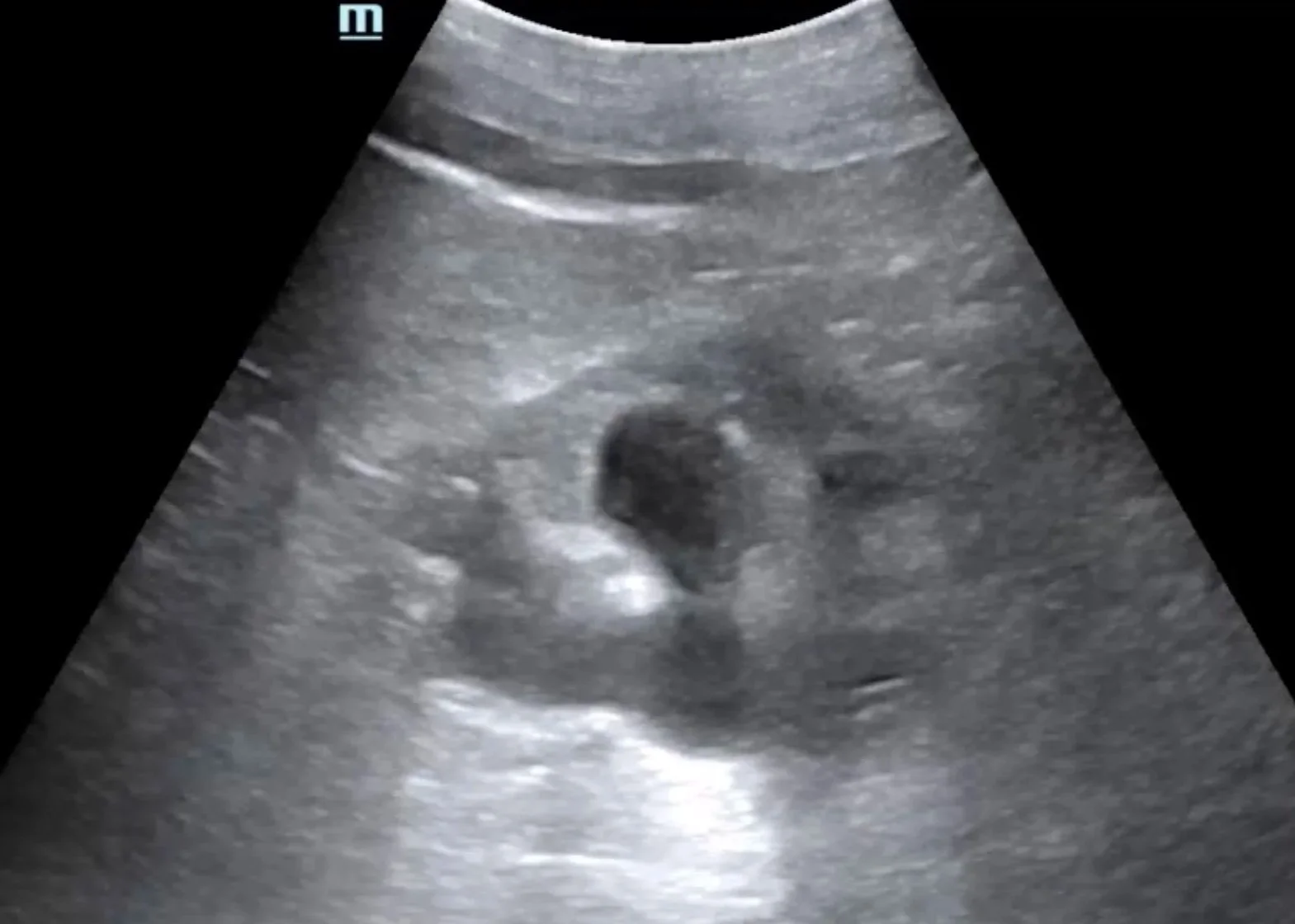
This case is by Dr. Nicole Minnerath and illustrates the detection of new hydronephrosis in a patient with bilateral nephrostomy tubes and ureteral stents. Read on to explore the role of renal and bladder POCUS in evaluating obstructive uropathy.
In this post, Dr. Tiana Sarsour highlights a case of recurrent abscesses and fever where POCUS confirmed a drainable collection, followed by a great review of abscess management and the role of ultrasound for skin and soft tissue infections.
This case is by Dr. Hannah Hendrix (now PGY2) and features a great example of the retrobulbar spot sign — a unique finding associated with embolic central retinal artery occlusion (CRAO). Read on for a quick review of ocular ultrasound and a closer look at this highly specific sonographic feature.
This post by Dr. Thomas Evans walks through the essentials of ultrasound-guided pericardiocentesis — when to do it, how to do it, and what pitfalls to avoid. We highlight the subxiphoid, parasternal, and apical approaches with practical POCUS tips for real-time needle visualization, plus a review of complications and how to keep the procedure as safe as possible.
To ring in our new year we wanted to spotlight another one of our former medical student rotators, Aubrey Strickland, who highlights the importance of using MAT in pregnancy. Remember narcan for everyone and to always call the poison center!
To get our medical student series started we want to introduce Student Doctor Janine Corley. She was inspired by our summer months in Cleveland and wanted to give a solid warning about eating shellfish. Enjoy this edition of Tox in the Land about Paralytic Shellfish Poisoning.
Lauren Porter DO, Ryan Marino MD, Bryan Ross MD, Marielle Brenner MD
Our latest blog post, by Sailakshmi Kumar, a fourth-year medical student at CWRU, explores ECMO in pregnancy with a focus on indications, physiological changes, and best practices for maternal and fetal care.
Welcome back to Tox in the Land!
After a short recess it’s time to get back at it. Since we are knee deep into interview season, there is no better time to put a spot light on our former chief resident and medical education fellow, Matthew Mullins, MD. He is here to guide you through the classic topic of iron toxicity and its management. Don’t worry, we will be back quickly to feature some of our medical student rotators as well!
Lauren Porter DO, Ryan Marino MD, Bryan Ross MD and Marielle Brenner, MD
This case is by Dr. Ryan Starkman (now PGY2) and is a great example of a large circumferential pericardial effusion with sonographic tamponade physiology diagnosed with POCUS. This expedited interventional cardiology involvement before the patient decompensated, and 750cc of fluid was successfully drained in the cath lab.
This case is by Dr. Jacob Perino and is a great example of an incidental finding of an iliac aneurysm found when performing a bladder ultrasound on a patient presenting with a malfunctioning Foley catheter. Read on to learn more about iliac aneurysms and bladder ultrasound in the setting of a Foley.
This month’s case is by Dr. Yanina Guevara who shares a case of painless monocular vision changes in a patient who was found to have a retinal detachment (and vitreous hemorrhage), first diagnosed with point-of-care ultrasound! This is accompanied by an overview of how to perform an ultrasound ocular exam and highlights some of the pathologies that can be visualized. Read on to learn more!