Intern Ultrasound of the Month: To Drain or Not to Drain? Ultrasound for Abscess
The Case
A 30-year-old male with a history of tricuspid valve endocarditis status post tricuspid valve replacement and prior IV drug use presented to the emergency department for fever and concern for abscess. He reported nearly a week of fevers, chills, nausea, painful swollen areas on his arm, and chest pain. He stated he has been abstinent from drugs for the past year but noted multiple prior abscesses requiring drainage.
He was mildly tachycardic with otherwise stable vitals. On exam, he had two raised, fluctuant areas. One lesion had an overlying eschar with mild surrounding erythema. No crepitus or drainage appreciated.
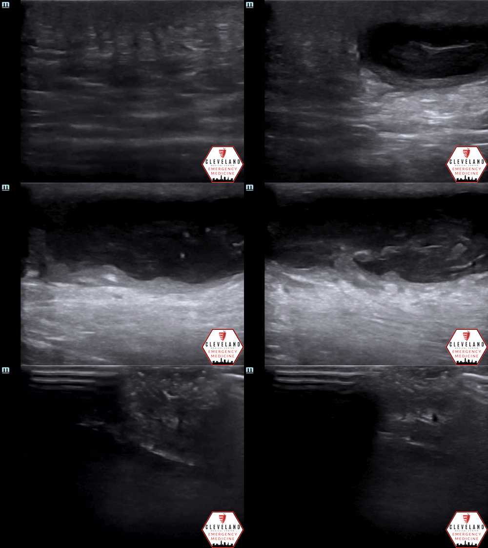
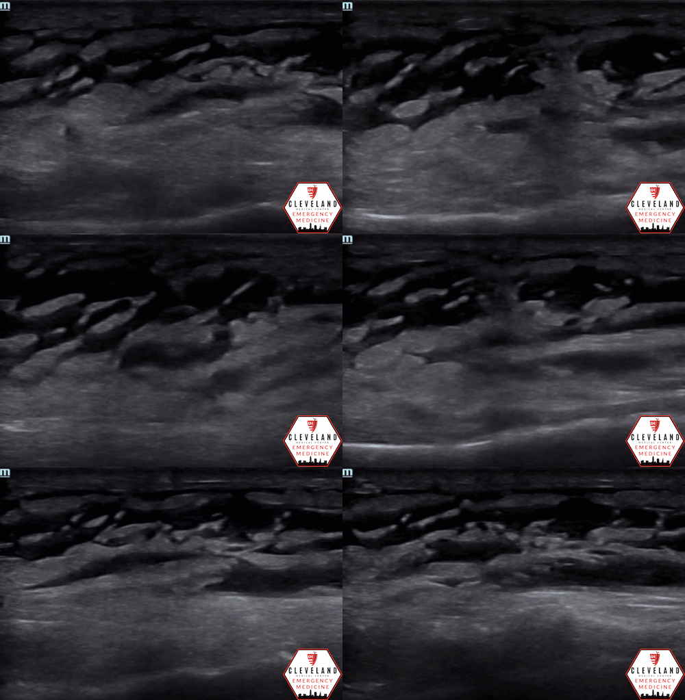
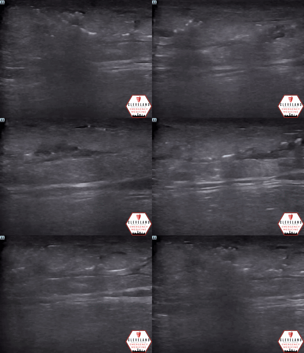
Point-of-care ultrasound (POCUS) was used to evaluate the lesions for abscess.
POCUS Findings
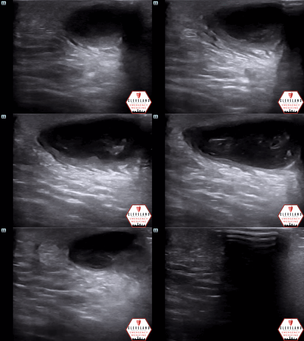
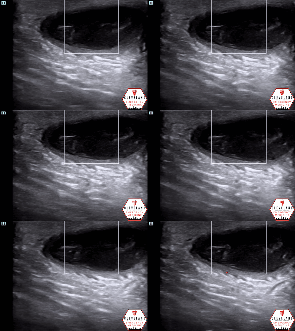
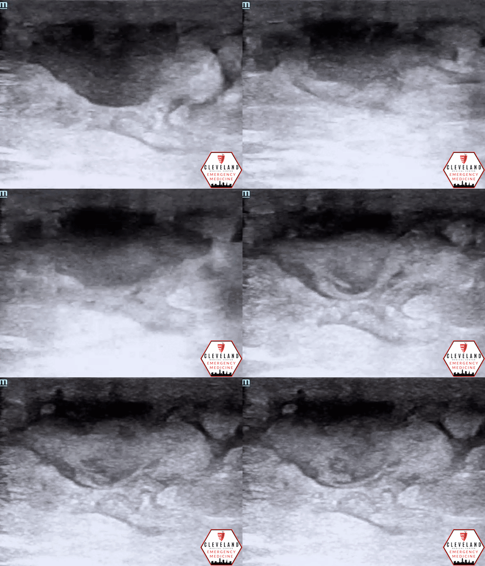
There is a well-circumscribed fluid collection with echogenic debris, consistent with an abscess. Surrounding cobblestoning was also present, supporting concurrent cellulitis in this clinical context. The second lesion demonstrated similar findings of a fluid collection with surrounding cellulitic changes.
Case Conclusion
The patient underwent bedside incision and drainage (I&D) and was started on broad-spectrum antibiotics given his history. He was admitted to the hospital for further management, and cultures ultimately grew methicillin-resistant Staphylococcus aureus (MRSA).
Abscess Formation
Overview
Skin and soft tissue infections (SSTIs) account for approximately 3% of all emergency department (ED) visits, representing more than three million visits per year [1]. An abscess is a localized collection of pus within the tissue, most often the result of a bacterial infection. Patients typically present with pain, tenderness, warmth, and swelling at the affected site, though deeper abscesses can also cause systemic symptoms such as fever, anorexia, and fatigue.
The process of abscess formation begins in compromised tissue where leukocytes accumulate. As neutrophils infiltrate the area, pus forms and necrosis of surrounding cells causes the cavity to expand. Risk factors for abscess formation include impaired host defenses, presence of foreign bodies, obstruction of normal drainage, and tissue ischemia or necrosis. If left untreated, abscesses can lead to complications such as bacteremic spread, rupture into adjacent structures, bleeding from erosion of nearby vessels, or impaired function of vital organs [2].
Microbiology
Most SSTIs are caused by Gram-positive organisms, most notably Staphylococcus aureus and Streptococcus pyogenes. S. aureus is particularly important given its ability to form abscesses and its variable resistance patterns, including methicillin-resistant strains (MRSA). In certain patient populations — such as those with recurrent infections, immunocompromise, or prior healthcare exposure — other pathogens may be implicated. These include enterococci, Gram-negative bacilli such as E. coli, and Pseudomonas aeruginosa, which may be associated with higher morbidity and mortality [3]. Recurrent abscesses should raise suspicion for MRSA, and empiric antibiotic coverage should reflect both common organisms and individual patient risk factors [4].
Diagnostics
Superficial abscesses can often be identified on physical exam by the presence of fluctuance, tenderness, and erythema. However, deeper collections may be more difficult to detect and often require imaging, such as CT or ultrasonography, for confirmation. POCUS can quickly identify a fluid collection, delineate its size and extent, and detect internal characteristics such as echogenic debris or loculations [5].
Another advantage of ultrasound is its ability to differentiate an abscess from cellulitis. Cellulitis typically demonstrates a cobblestone pattern of hyperechoic fat lobules separated by hypoechoic fluid, while abscesses appear as discrete fluid collections with well-defined borders. Ultrasound can also identify nearby vasculature with the use of color Doppler, reducing the risk of complications I&D. In some cases, POCUS has been shown to change management in nearly half of patients initially thought to have cellulitis [6].
Management
When to drain?
The primary treatment for most abscesses is I&D, which can usually be performed safely at the bedside. Smaller abscesses (< 1cm) may resolve with warm compresses and close follow-up, while larger or deeper collections sometimes require image-guided aspiration or surgical consultation [3].
When are antibiotics indicated?
Antibiotics are not always required after drainage, but they are indicated for patients with multiple or recurrent abscesses, significant cellulitis, systemic illness, immunocompromise, or abscesses greater than 2 cm [4]. Empiric therapy should target the most likely pathogens: first-generation cephalosporins or anti-staphylococcal penicillins are appropriate for many community-acquired infections. Outpatient options with MRSA coverage include trimethoprim-sulfamethoxazole, doxycycline, or clindamycin, while cephalexin or dicloxacillin remain appropriate for non-purulent cellulitis. Severe or complicated infections may require treatment with vancomycin or other advanced agents, with broader regimens such as vancomycin plus piperacillin-tazobactam for high-risk or polymicrobial cases [7].
Importantly, recurrent abscesses should raise suspicion for MRSA, and cultures should be obtained whenever possible to guide therapy [4].
Ultrasound Technique for Identifying Abscess
Probe selection: Use a linear transducer for most soft tissue abscesses; switch to a curvilinear probe if the collection is large or deep.
Establish normal: Begin scanning away from the affected area to appreciate normal soft tissue appearance, see Figure 1.
Assess the affected area: Slide or fan the probe across the area (from one end to the other), then rotate 90° to obtain orthogonal views and define its full extent.
Color Doppler: If a fluid collection is visualized, apply color doppler to identify nearby vasculature and differentiate abscess from pseudoaneurysm [8].
Correlate anatomy: Place a fingertip on the area of maximal fluctuance, then slide the probe to the fingertip to align surface anatomy with the ultrasound image.
Guidance for drainage: Consider real-time ultrasound guidance during I&D, regardless of size or depth, to improve accuracy and safety.
Post-procedure check: Repeat ultrasound after drainage to assess for residual fluid [9].
Figure 1. Normal skin, subcutaneous tissue, and fascia [8].
Ultrasound Findings
Figure 2. Abscess with echogenic debris, swirling with compression.
Abscess
Well-circumscribed, anechoic or hypoechoic fluid collection.
Often contains hyperechoic debris from pus or necrotic material.
Walls are usually distinct and hyperechoic.
Probe compression may produce swirling of fluid, known as the “squish sign” (see Figure 2) [8].
Cellulitis
Early: thickened skin and increased echogenicity with loss of distinct soft tissue layers (see Figure 3).
Advanced: hypoechoic fluid tracking between fat lobules, producing the classic cobblestone appearance, as seen in Figure 4 [5].
May occur with or without an associated abscess.
Figure 3. Early cellulitis (left) versus normal [8]
Figure 4. Late cellulitis with cobblestoning
*Note: Cobblestoning and well-circumscribed fluid collections are not specific for infection and may also be seen in non-infectious processes such as hematomas or sterile fluid collections.
Complicated infections
Ultrasound may reveal fluid along fascial planes or subcutaneous air (Figures 5 and 6, respectively), raising concern for necrotizing fasciitis [9].
Figure 5. Fluid tracking along the fascial plane, suggesting early necrotizing fasciitis
Figure 6. Subcutaneous air in the soft tissues, a late finding in necrotizing fasciitis
Utility of POCUS for Abscess
POCUS improves patient care by helping clinicians distinguish cellulitis from abscess and determine the extent. This ensures that patients with drainable fluid collection receive timely I&D, while those without abscess avoid unnecessary procedures [5].
Evidence suggests that ultrasound frequently changes management decisions in suspected cellulitis. In one prospective study, it altered management in 39 of 82 patients (48%): 33 went on to receive drainage, while 6 were directed toward further diagnostics or consultation. Among those in whom drainage was originally believed to be needed, ultrasound changed management in 32 of 44 patients (73%) — eliminating drainage in 16 and prompting additional diagnostics in 16 others [6]. In a randomized controlled trial, patients who underwent I&D with POCUS had significantly lower treatment failure rates (defined as the need for repeat drainage or ongoing infection) compared with those managed by physical exam alone [10].
POCUS can also detect concerning findings such as fluid tracking along fascial planes or subcutaneous emphysema, which should raise immediate concern for necrotizing fasciitis, a surgical emergency requiring prompt recognition and intervention [9].
Take-Home Points
SSTIs are common in the ED, and ultrasound is a powerful adjunct for evaluation.
POCUS can differentiate cellulitis from abscess and guide safe drainage.
Dynamic ultrasound guidance with color Doppler improves safety and helps confirm adequate drainage.
Recurrent abscesses should raise suspicion for MRSA, and cultures are essential for guiding therapy.
Ultrasound frequently changes management decisions and improves patient outcomes.
AUTHORED BY: TIANA SARSOUR, MD
FACULTY CO-AUTHOR/EDITOR: LAUREN MCCAFFERTY, MD
References
Yusuf S, Hagan JL, Adekunle-Ojo AO. Managing Skin and Soft Tissue Infections in the Emergency Department Observation Unit. Pediatr Emerg Care. 2019;35(3):204-208.
Bush LM. Abscesses – Infectious Diseases. Merck Manuals Professional Edition. December 8, 2023. Accessed January 30, 2024. https://www.merckmanuals.com/professional/infectious-diseases/biology-of-infectious-disease/abscesses
Ioannou P, et al. Gram-Negative Bacteria as Emerging Pathogens Affecting Mortality in Skin and Soft Tissue Infections. Hippokratia. 2018;22(4):153-161. https://www.ncbi.nlm.nih.gov/pmc/articles/PMC6528699/
Stevens DL, Bisno AL, Chambers HF, et al. Practice Guidelines for the Diagnosis and Management of Skin and Soft Tissue Infections: 2014 Update by the Infectious Diseases Society of America. Clin Infect Dis. 2014;59(2):e10-e52.
O’Rourke K, et al. Ultrasound for the Evaluation of Skin and Soft Tissue Infections. Mo Med. 2015;112(3):202-205. https://www.ncbi.nlm.nih.gov/pmc/articles/PMC6170135/
Tayal VS, et al. The Effect of Soft Tissue Ultrasound on the Management of Cellulitis in the Emergency Department. Acad Emerg Med. 2006;13(4):384-388.
Fung HB, et al. A Practical Guide to the Treatment of Complicated Skin and Soft Tissue Infections. Drugs. 2003;63(14):1459-1480.
American College of Emergency Physicians. Abscess Evaluation. ACEP Sonoguide. Accessed January 30, 2024. https://www.acep.org/sonoguide/procedures/abscess-evaluation
Menegas S, et al. Abscess Management: An Evidence-Based Review for Emergency Medicine Clinicians. J Emerg Med. 2020;59(6):817-828.
Gaspari RJ, Sanseverino A, Gleeson T. Abscess Incision and Drainage With or Without Ultrasonography: A Randomized Controlled Trial. Ann Emerg Med. 2019 Jan;73(1):1-7.
