Intern Ultrasound of the Month: A Sign of Acute Pulmonary Embolism
The Case
77-year-old female with past medical history pertinent for end-stage renal disease, heart failure, and lung cancer who presented to the emergency department (ED) after becoming unresponsive. The patient's family reported that she had been in her usual state of health earlier that day, but while sitting in a chair she suddenly slumped over and stopped responding. EMS arrived, and she was awake and talking en route to the ED. Shortly after arrival, she again became unresponsive. She was found to be pulseless, with pulseless electrical activity (PEA) on the monitor, and ACLS protocol was started. During the resuscitation, a cardiac point-of-care ultrasound was performed to evaluate for potential causes of her PEA arrest.

POCUS Findings/Interpretation
An apical 4 chamber image series obtained during a brief period of return of spontaneous circulation (ROSC) shows a significantly enlarged right ventricle with free wall hypokinesis and apical sparing, otherwise known as McConnell's Sign. This was concerning for PE.

CT demonstrating saddle PE
Case Conclusion:
The patient went into PEA arrest again. Family continued to want to pursue aggressive measures. No other reversible causes were identified. Given her acute onset of symptoms, cancer history, and ultrasound findings, PE was a major concern. Benefits and risks of thrombolytics were considered, and in the setting of recurrent cardiac arrest, the decision to push IV tPA was made. Following tPA administration, ROSC was achieved. Once stabilized, CT was performed which showed a saddle PE. She was transferred to the ICU for further management.
Acute Pulmonary Embolism
Acute pulmonary embolism (PE) is a common clinical entity, with an incidence ranging from 39 to 115 per 100,000 population annually (1). The presentation of patients with PE has an extensive range of clinical implications and severity. PE can be asymptomatic and identified incidentally on imaging studies but, as seen in this case, can traverse the entire spectrum of severity and present as cardiac arrest.
A PE is a disruption or total blockage of flow of blood in the pulmonary arterial tree. These blockages are typically caused by thrombi, most often deep venous thrombosis (DVTs) of the lower extremities (2). PE can also arise in situations where materials other than blood, such as fat, air, or foreign bodies, migrate to the pulmonary arterial system, although this is exceedingly rare when compared with DVT (3).
The classification of PE is based not on the size of the clot or clot burden but the hemodynamic instability that may result as a function of obstructive shock. PEs that cause hemodynamic instability should be referred to and documented as such. This is defined by a systolic blood pressure (SBP) less than 90 mmHg, a drop in SBP by greater than 40mm Hg from baseline, or hypotension requiring vasopressor support; such PEs were previously referred to as 'massive' or 'high-risk'. Hemodynamically stable PEs can range from small, incidentally found, and asymptomatic PE to those which may cause initial hypotension that is fluid-responsive or those causing right ventricular (RV) dysfunction without hemodynamic instability. These are referred to as "low-risk” or ”small” PEs and "sub-massive” or ”intermediate-risk” PEs, respectively (4).
Computed tomography (CT) angiography is the preferred imaging study for PE. While generally not diagnostic of PE, transthoracic echocardiography can detect signs of right heart strain, which can support the diagnosis of PE in the right clinical context (4-7). This can be especially helpful in situations in which patients are too unstable for CT, there is a contraindication to CT, or other confirmatory tests are not readily available.
Given the wide variety of clinical presentation, treatment for PE varies greatly as well. Treatment ranges from anticoagulation only to systemic thrombolytic administration, catheter-directed therapy, or surgical embolectomy (4).
McConnell’s Sign on Point-of-Care Ultrasound

Figure 1. McConnell’s sign - hypokinesis of the RV free wall with preserved apical contractility.
McConnell’s Sign & Its Significance
This ultrasound finding of regional right ventricular dysfunction, with akinesia of the mid free wall and preserved contractility of the apical wall, is known as McConnell's sign (5), see Figure 1. This is a poorly sensitive but highly specific and early indicator for acute PE and has particular diagnostic utility in patients who are too unstable for CT or are being considered for thrombolytic administration (6-7).
McConnell's sign, while highly specific, has occasionally been seen in cases of right ventricular ischemia in the absence of PE (8). However, the rarity of these "PE-less McConnell's signs" is such that any patient with an identified McConnell's sign on ultrasound should be strongly considered for acute PE.
McConnell's sign is best appreciated in the apical four chamber view as it allows for visualization of the RV free wall, tricuspid annulus, and apex.
Image Acquisition
Acquiring the apical four chamber view can be difficult, especially in cases such as ours where the patient had an automated CPR device on the chest. Difficulties also arise with body habitus, respiratory distress, and patient positioning. In general, a good first step in your approach to obtaining the desired view is to place the transducer in the 4th or 5th intercostal space often near or lateral to the nipple line with the probe marker facing the 2-3 o’clock position (in cardiac convention), see Figures 2 and 3. If patient condition permits, the left lateral decubitus position can be employed to more readily obtain the desired sonographic windows, as this will bring the heart closer to the chest wall. In an adequate view, all four cardiac chambers are visualized. In addition, the interventricular septum should be vertically oriented on your screen (which often requires rocking the probe toward the axilla), as this will give you the most accurate assessment (9-11); see Figure 4.

Figure 2. Probe placement for apical four chamber view. Image credit: ACEP Sonoguide (10).

Figure 3. Probe placement. Image credit: IMPoCUS (11)

Figure 4. Apical 4 chamber view
Other Sonographic Signs of Right Heart Strain
In addition to McConnell’s sign, other key sonographic signs of right heart strain include:
Right ventricular (RV) enlargement - Normally, the RV to LV ratio is roughly 0.67. When the RV approaches or exceeds the size of the LV, this is considered pathologically enlarged. This can best evaluated in the apical four chamber view (Figure 5).
Abnormal septal motion (“D sign”) - When the right ventricle bows towards the left this indicates increased right-sided pressures. This appears as a “D-sign” on a parasternal short axis view, as shown in Figures 6 and 7.
Tricuspid annular plane systolic excursion (TAPSE) - Briefly, this is a way to estimate RV contractility. Because the RV primarily contracts longitudinally (compared to the LV which contracts concentrically), vertical displacement of the tricuspid annulus correlates with RV contraction. Therefore, decreased TAPSE (i.e. decreased vertical displacement) indicates reduced contractility. TAPSE less than 17 mm is considered abnormal (11-13)
How to do it: Obtain an apical four chamber view. Place m-mode cursor over the lateral tricuspid valve annulus. The resulting waveform shows the displacement of tricuspid annulus through the cardiac cycle. Measure the vertical height between peak and trough. See Figure 8.
There are additional, more complex methods to evaluate for right heart strain using ultrasound. See resources below for more details.

Figure 5. Apical four chamber view demonstrating RV enlargement. Note that all four cardiac chambers are visualized, the septum is vertical, and you can clearly see the RV free wall.

Figure 6. Parasternal short axis view demonstrating septal flattening, resulting in the LV having a “D” shape.
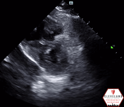
Figure 7. Parasternal short axis view from our patient, demonstrating septal flattening

Figure 8. TAPSE measurement using M-mode
AUTHORED BY: GABE ALAGNA, MD (PGY1)
FACULTY CO-AUTHOR/EDITOR: LAUREN MCCAFFERTY, MD
References
Wendelboe AM, Raskob GE. Global Burden of Thrombosis: Epidemiologic Aspects. Circ Res. 2016;118(9):1340-7.
Turetz M, Sideris AT, Friedman OA, Triphathi N, Horowitz JM. Epidemiology, Pathophysiology, and Natural History of Pulmonary Embolism. Semin Intervent Radiol. 2018;35(2):92-98
Khashper A, Discepola F, Kosiuk J, Qanadli SD, Mesurolle B. Nonthrombotic pulmonary embolism. Am J Roentgenol. 2012;198(2):W152-9
Vyas V, Goyal A. Acute Pulmonary Embolism. [Updated 2022 Aug 8]. In: StatPearls [Internet]. Treasure Island (FL): StatPearls Publishing; 2024 Jan. Available from: https://www.ncbi.nlm.nih.gov/books/NBK560551/
McConnell MV, Solomon SD, Rayan ME, Come PC, Goldhaber SZ, Lee RT. Regional right ventricular dysfunction detected by echocardiography in acute pulmonary embolism. Am J Cardiol. 1996;78:469–473.
Patra S, Math RS, Shankarappa RK, Agrawal N. McConnell's sign: an early and specific indicator of acute pulmonary embolism. BMJ Case Rep. 2014;2014:bcr2013200799
Fields JM, Davis J, Girson L, Au A, Potts J, Morgan CJ, Vetter I, Riesenberg LA. Transthoracic Echocardiography for Diagnosing Pulmonary Embolism: A Systematic Review and Meta-Analysis. J Am Soc Echocardiogr. 2017;30(7):714-723.e4
Rafie N, Foley DA, Ripoll JG, Booth-Kowalczyk ML, Arghami A, Pochettino A, Michelena HI. McConnell's Sign Is Not Always Pulmonary Embolism: The Importance of Right Ventricular Ischemia. JACC Case Rep. 2022;4(13):802-807.
Koratala A. Focused Cardiac Ultrasound for the Nephrologist: The apical window. Renal Fellow Network. September 20, 2019. Retrieved from: https://www.renalfellow.org/2019/09/20/focused-cardiac-ultrasound-for-the-nephrologist-the-apical-window.
5 Core Views. IMPoCUS. Retrieved from: https://www.impocus.ca/cardiac-focus---5-core-view---5
Prats MI, Bahner DP. Cardiac. Sonoguide. August 18, 2020 Retrieved from: https://www.acep.org/sonoguide/basic/cardiac.
Lang RM, Badano LP, Mor-Avi V, et al. Recommendations for cardiac chamber quantification by echocardiography in adults: an update from the American Society of Echocardiography and the European Association of Cardiovascular Imaging. J Am Soc Echocardiogr. 2015;28(1):1-39.e14
Alerhand S, Sundaram T, Gottlieb M. What are the echocardiographic findings of acute right ventricular strain that suggest pulmonary embolism? Anaesth Crit Care Pain Med. 2021;40(2):100852