Intern Ultrasound of the Month: Retinal Detachment
The Case
75-year-old male with past medical history significant for pituitary adenoma (status post resection) who presented with a 24-hour history of acute painless vision loss in his left eye. This started suddenly while riding in a car when he developed a “black hole” in the inferomedial aspect of his left eye. He then felt a “curtain closing down over his eye” with decreased vision in the lower visual fields of that eye. He reported having a history of fuzzy vision in his left eye, occasional floaters, and dry eyes but felt that presenting symptoms were different. He denied any discharge from his eye, periorbital edema, or fevers but did report that his eye felt a little itchy. He also denied any headache, dizziness, or weakness or numbness in the upper or lower extremities.
Ocular point-of-care ultrasound (POCUS) was performed to evaluate for retinal detachment.


POCUS Findings:
There is a large lateral retinal detachment indicated by a distinct hyperechoic linear structure in the posterior chamber that has separated from the choroid and is tethered on temporal side of the vitreous body (screen right). This is further appreciated with the kinetic exam, in which the patient looked left and right. There is also a highly echogenic structure at the base of the optic nerve, most likely drusen, which are calcium deposits that are usually benign.
Case Continued: Ophthalmology was consulted, and through their comprehensive assessment they confirmed retinal detachment with macular involvement (“mac-off”). The patient was discharged with plan to return 8 hours later for laser intervention and reattachment of the detached portion of the retina with a retina specialist.

Figure 1. Retinal Detachment
Source: https://www.mayoclinic.org/diseases-conditions/retinal-detachment/symptoms-causes/syc-20351344
Retinal Detachment
Pathophysiology of Retinal Detachment
Through the aging process, the vitreous humor shrinks and thins. The vitreous humor moves, without friction, around on the retina. However, if a portion of the vitreous sticks to the retina, it can pull on the attached portion causing a tear allowing fluid to pass through and lift the retina [1]. It is important to remember that the retina is a continuation of the optic nerve. Conditions that increase likelihood of developing a retinal detachment are severe myopia, prior eye surgery (cataract, glaucoma), eye trauma, prior retinal detachments. Common signs and symptoms of retinal detachment are sudden onset of floaters or flashers, shadowing or a “curtain” over the field of vision. [1].
Anywhere from 1.3-3% of emergency department visits are due to eye-related complaints [2-3]. While retinal detachment is not one of the more common pathologies, it is considered an eye emergency for which prompt recognition is important [4].
Review of Imaging Modalities for Retinal Detachment
There are several imaging options, including ultrasound, CT, or MRI, that can be used to evaluate eye conditions. Depending on the pathology, these have varying diagnostic utility [3, 5-7]. While the gold standard for diagnosing retinal detachment is a dilated fundoscopic examination, performed primarily by an ophthalmologist, ultrasound has been used by ophthalmologists for decades. More recently emergency physicians have utilized POCUS to evaluate for retinal detachment and have demonstrated a sensitivity and specificity of 97% -100% and 83% -100%, respectively [3]. Another study among a large heterogeneous group of emergency physicians showed sensitivity of 75% and specificity of 94% [5]. MRI is generally not the preferred imaging modality for evaluating retinal detachment as time to acquire image is lengthy, it is expensive, and may provide a limited assessment. Also, if there is a possibility of metallic foreign body in the globe, MRI would not be favorable. MRI is useful in evaluating for the underlying cause of retinal detachment such as a mass, disease within the globe and orbit, or optic nerve pathology [6]. Given cost, timing, and other logistical barriers associated with MRI, POCUS is a fast and reliable diagnostic modality that should be incorporated into the standard initial workup for retinal detachment, along with ophthalmologist exam, unless there is concern for an open globe injury. It is a particularly useful option when ophthalmology is not immediately available or if a fundoscopic exam cannot be performed [8].
Materials:
High frequency linear probe
Tegaderm to cover eye
Ultrasound gel
Techniques for Ocular Ultrasound

Figure 2. Probe placement [9]
Place a transparent adhesive covering, such as a tegaderm, over the patient’s eye and ensure there are no air pockets. Using a linear transducer (10-5 MHz) offers the highest resolution given the superficial depth of the structures of the eye. The technician should stabilize their hand gently on the patient’s nasal bridge or zygoma to prevent excessive force on the eye with the probe. Probe marker should face the patient’s right side.
With a normal gain setting, adjust depth to allow for visualization of the posterior segment of the eye that includes the retina and optic nerve. Identify (from top to bottom) the cornea, anterior chamber, lens, vitreous chamber, retina, and optic nerve (see Figure 3). Once these structures are identified, reduce gain until the vitreous humor is anechoic. Then using oculokinetic echography, i.e. having the patient look left and right and evaluating the dynamic movement of the eye with ultrasound, examine the vitreous humor for any abnormalities that are not firmly attached to the retina [9-10].

Figure 3. Probe placement and eye anatomy [10]
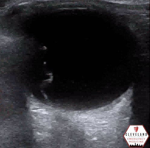
Figure 4. Retinal detachment
Retinal Detachment
Often visualized in normal or low gain settings, retinal detachment appears as a hyperechoic mobile cord-like structure that is attached to or near the optic nerve in the posterior wall of the eye. If the retinal detachment is large, the flap is often easily visualized where it attaches to the optic nerve sheath. For smaller retinal detachment or those not near the optic nerve, scan through the eye horizontally and vertically and then slow down the image after capture to carefully evaluate for a tethered flap. In addition to its relation to the optic nerve, retinal detachment tends to be less mobile with kinetic movement compared to vitreous detachment or hemorrhage [9-10].
Vitreous Detachment & Hemorrhage
Best visualized with high gain settings, both tend to be more free floating with a “washing machine” appearance with kinetic movement in the posterior chamber of the eye. Posterior vitreous detachment (PVD) often has a thin hyperechoic flap that crosses over the optic nerve and is not tethered to the posterior wall. Vitreous hemorrhage (VH) appears as diffuse echogenic material and may have a “snowglobe” appearance. It is frequently seen in diabetic patients, is easier to distinguish from posterior vitreous detachment and retinal detachment, and is not an ocular emergency [9-10].


Take Home Points
Retinal detachments make up a small percentage of atraumatic eye presentations but require emergent evaluation for definitive treatment. Conditions associated with risk of developing RD are severe myopia, prior eye surgery, eye trauma, prior retinal detachments
Retinal detachment appears as a hyperechoic flap within the vitreous body that is tethered to the posterior wall at or near the optic nerve. Use oculokinetic echography and slow down image to optimize your evaluation
Slowly increase the gain to evaluate for PVD and VH, which are more free floating (washing machine sign) and can cross midline with no relation to the optic nerve.
POST BY: DR. RANJANA RAVIKUMAR, PGY1
FACULTY CO-AUTHOR/EDITOR: LAUREN MCCAFFERTY, MD
References
Boyd K. Detached retina. American Academy of Ophthalmology. October 13, 2022. Retrieved May 1, 2023. <https://www.aao.org/eye-health/diseases/detached-torn-retina>
Nash EA, Margo CE. Patterns of emergency department visits for disorders of the eye and ocular adnexa. Arch of Ophthalmol. 1998;116(9):1222–1226.
Vrablik ME, Snead GR, Minnigan HJ, Kirschner JM, Emmett TW, Seupaul RA. The diagnostic accuracy of bedside ocular ultrasonography for the diagnosis of retinal detachment: A systematic review and meta-analysis. Annals Emerg Med. 2018; 65(2).
Channa R. Zafar SN, Canner JK, Haring RS, Schneider EB, Friedman DS. Epidemiology of eye-related emergency department visits. JAMA Ophthalmol. 2016; 134(3):312-9.
Kim DJ, Francispragasam M, Docherty G., Silver B., Prager R, Lee D, Maberley D. Test characteristics of point‐of‐care ultrasound for the diagnosis of retinal detachment in the emergency department. Acad Emerg Med. 2019;26(1):16-22.
Hallinan JT, Pillay P, Koh LH, Goh KY, Yu WY. Eye Globe Abnormalities on MR and CT in Adults: An Anatomical Approach. Korean J Radiol. 2016 Sep-Oct;17(5):664-73. doi: 10.3348/kjr.2016.17.5.664. Epub 2016 Aug 23. PMID: 27587955; PMCID: PMC5007393.
Skidmore C, Saurey T, Ferre RM, Rodriguez-Brizuela R, Spaulding J, Lundgreen Mason N. A narrative review of common uses of ophthalmic ultrasound in emergency medicine. J Emerg Med. 2021; 60(1), 80–89.
Botwin A, Engel A, Wasyliw C. The use of ocular ultrasound to diagnose retinal detachment: A case demonstrating the sonographic findings. Emerg Radiol. 2018; 25(4), 445–447.
Nagdev A. Ocular ultrasound: Retinal detachment and Posterior Vitreous Detachment. ALiEM. March 11, 2014. Retrieved April 20, 2023. <https://www.aliem.com/ocular-ultrasound-retinal-detachment-posterior-vitreous-detachment/>
Dinh V. Ocular Ultrasound made easy: Step-by-step guide. POCUS 101. Accessed May 1, 2023. <https://www.pocus101.com/ocular-ultrasound-made-easy-step-by-step-guide/>