Intern Ultrasound of the Month: Pneumothorax
The Case
65-year-old male with a past medical history of hypertension and hyperlipidemia presented to the emergency department by EMS following a MVA in which he was the restrained driver. The patient stated that the last thing he remembered was seeing his “check engine light” turned on so he pulled off the road and apparently struck a concrete barricade. He reported loss of consciousness after the accident with poor recollection of events surrounding the accident. Per EMS, the patient's car was found to have significant front end damage and airbag deployment. The patient had self-extricated and EMS found him near his vehicle reporting right leg/ankle pain, which he described as a ”7 out of 10” in severity. Patient otherwise denied any chest pain or shortness of breath.
On arrival to ED, his vital signs were: temperature 36.5, blood pressure 123/91, heart rate 85, respiratory rate of 20, SPO2 90% on room air.
Physical exam was notable for seatbelt sign across the abdomen and chest, diminished breath sounds in the right upper lobe, and visible swelling, ecchymoses, deformity of the right ankle with tenderness to palpation but neurovascularly intact.
The patient was placed on 4L nasal cannula with improvement in oxygenation. An eFAST exam was performed to evaluate for pneumothorax as well as hemopericardium/hemoperitoneum.

Lack of lung sliding in the least dependent area of the lung

Lung point, indicating the presence of a pneumothorax
POCUS findings: When the linear probe was placed over the right third and fourth ribs (with the probe indicator pointing cephalad), there was a lack of lung sliding. As the probe was moved slightly laterally and more caudally, a lung point was seen. Not shown here but lung sliding was observed in the basilar segments of the right lung and throughout the left lung. The remainder of the eFAST views revealed no evidence of hemothorax, hemoperitoneum, or hemopericardium.
Impression: lung ultrasound significant for right-sided pneumothorax
Case Continued: Chest x-ray confirmed pneumothorax. A chest tube was placed with subsequent improvement in respiratory status. CT imaging revealed displaced fractures of right anterolateral 2nd-4th ribs. The chest tube was in place and only a small residual pneumothorax was noted. Additional injuries included a bimalleolar fracture and tibial plateau fracture. The patient had a 14-day hospital course during which the extremity fractures were operatively managed. The chest tube was removed on day 4, and the patient was ultimately discharged to a rehab facility for further care.
Pneumothorax
Epidemiology/Etiology [1,2]
Can be spontaneous (primary vs secondary) or traumatic/iatrogenic
Most commonly traumatic/iatrogenic with incidence of 13.9/100,000 in the US population.
Cost per case estimated to be $20,000-$30,000
Risk factors for spontaneous pneumothorax include smoking and male gender
Clinical signs and symptoms [2,3]
Ipsilateral chest pain, often pleuritic
Acute dyspnea
May have normal vitals and exam in cases of a small pneumothorax (<15% hemithorax)
For a larger pneumothorax, there may be absent or diminished breath sounds and tactile fremitus as well as hyperresonant percussion
Hypotension, tachypnea, significant tachycardia should raise suspicion for tension pneumothorax
Diagnostic imaging [2,4-6]
CT
“Gold standard”
Cons:
Costly
Time consuming
Inappropriate for unstable patients
Radiation exposure
CXR
Best when patient is upright and films are PA
Cons:
Low sensitivity
Radiation exposure
Ultrasound
Significantly higher sensitivity (88%) compared to CXR (52%) with similar specificity (99-100%) [5]
Pros:
Portable
Patient can be assessed in supine position (a common position of trauma patients)
No radiation exposure
Faster diagnosis than CT or CXR [6]
Cons:
Operator-dependent
See pitfalls below
Management [7]
Conservative management if small and stable (oxygen, re-image)
Tube thoracotomy
Pigtail/small bore catheter
Large bore chest tube if concerned about hemothorax
Video-assisted thoracoscopic surgery (VATS) is an option
POCUS Assessment for Pneumothorax [4,8-11]
Indications
Absent or abnormal breath sounds on pulmonary auscultation
Risk factors for pneumothorax including blunt or penetrating chest trauma
Clinical signs or symptoms of pneumothorax such as hypoxia or respiratory distress

Technique
Probe: High frequency (5-10 MMHz) linear probe is best although curvilinear prove may also be used
Patient positioning & probe placement — focus is on the least dependent area of the lung
Supine
Place transducer longitudinally (indicator pointing toward patient’s head) over the anterior chest, around the level of ribs 3rd-4th intercostal space
Upright (less sensitive)
Place probe longitudinally at the apex of lungs. Note that vertical movement with respiration is less pronounced as in lower fields.
Will need to scan up and down a rib space to ensure full evaluation of the least dependent region
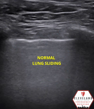
Keeping the probe perpendicular to the chest wall, identify the pleural line (which is the hyperechoic line slightly deep to the hyperechoic arc of the ribs)

In a normal lung, the pleural line will exhibit lung sliding, the horizontal back-and-forth movement resulting from the parietal and visceral pleura sliding over one another.
Findings suggestive of pneumothorax
Lack of lung sliding and comet tail artifacts may be seen with a pneumothorax but this is nonspecific! May occur in a number of other conditions including bullae, adhesive lung disease, prior pleurodesis, etc. Clinical context is key!
In M-mode, will see two distinct patterns on M mode (Sandy Beach vs. Barcode sign)


https://www.grepmed.com/images/4336/stratosphere-pocus-diagnosis-seashore-pneumothorax
Lung point is highly specific for pneumothorax — the point at which the visceral and parietal pleura detach (absent sliding is immediately adjacent to sliding)
Location of lung point can estimate size (the more lateral and caudal, generally the larger the size)
Can monitor this over time to determine improvement vs worsening


Image from: https://litfl.com/lung-ultrasound-lung-point/
What rules out a pneumothorax?
Lung sliding (in the least dependent area of the lung) - see normal above
B lines/comet-tail artifacts*
Lung pulse* — subtle pulsating-like movement of the pleural line resulting from cardiac motion transmitted through the lung parenchyma
*requires direct contact between the parietal and visceral pleura in order to be seen. Even if lung sliding is absent, these findings also rule out a PTX

Pitfalls
False positive lack of lung sliding: misidentifying the pleural line — rib, fascial planes, etc. can be mistaken for the pleural line and won’t exhibit lung sliding
Large pleural effusion and subcutaneous emphysema can inhibit visualization of the pleural line
A lung point may not be visualized in a massive pneumothorax
False positive lung point: interface of the lung and diaphragm/heart/abdominal organ can be mistaken for a lung point (be mindful of this when scanning low on the chest or over the heart)
Look at the structures deep to what appears to be the pleural line. If you see movement, tissue, and absence of a-lines, this is most likely not a true lung point

Source: https://www.coreultrasound.com/uotw-62-answer/
Take Home Points
Lung ultrasound is a great bedside tool to quickly evaluate for pneumothorax (among other conditions). While it’s limited by operator experience, POCUS has demonstrated significantly higher sensitivity compared to chest x-ray and has advantages over CT.
The presence of lung sliding and/or comet tail artifacts/B lines rules out a pneumothorax while a lung point rules in a pneumothorax
AUTHORED BY: DR. BLAKE NELSON (R1)
FACULTY CO-AUTHOR/EDITOR: LAUREN MCCAFFERTY, MD
References
Srinivas R. Mummadi JK. Epidemiology of Adult Pleural Disease in the United States. Chest. 2021; 160(4):1534-1551.
Noppen M, De Keukeleire T. Pneumothorax. Respiration. 2008; 76: 121-127.
Sahn, S. A., & Heffner, J. E. (2000). Spontaneous pneumothorax. N Engl J Med. 342(12), 868-874.
Staub LJ, Biscaro RR, Kaszubowski E, Maurici R. Chest ultrasonography for the emergency diagnosis of traumatic pneumothorax and haemothorax: A systematic review and meta-analysis. Injury. 2018; 49: 457-466.
Ding W, Shen Y, Yang J, He X, Zhang M. Diagnosis of pneumothorax by radiography and ultrasonography: A meta-analysis. Chest. 2011; 140(4): 859–866.
Zhang M, Zhi-Hai L, Yang YX, Gan JX, Xu SW, You XD, Jiang GY. Rapid detection of pneumothorax by ultrasonography in patients with multiple trauma. Crit Care; 10(4): 112.
DeMaio A, Semaan R. Management of Pneumothorax. Clin Chest Med. 2021;42(4):729-738.
Ma JO, Mateer JR, Kirkpatrick AW. Trauma. In OJ Ma, JR Mateer, RF Reardon, SA Joing (eds), Ma and Mateer’s Emergency Ultrasound (3rd ed). 2008. New York, NY: McGraw-Hill Education. pp 61-92.
Noble V, Nelson B. Focused assessment with sonography in trauma (FAST). In Manual of Emergency and Critical Care Ultrasound. 2011. Cambridge: Cambridge University Press. pp. 27-60.
Alerhand S, Gulalp B. Lung. ACEP Sonoguide. 2021 Mar 8. Retrieved April 2022 from https://www.acep.org/sonoguide/basic/lung/
Smith B. “USOTW #62.” Core Ultrasound. 2015 Oct 25. Retrieved April 2022 from <https://www.coreultrasound.com/uotw-62-answer/