Intern Ultrasound of the Month: A Mimicker of Vitreous Hemorrhage - Asteroid Hyalosis
THE CASE
60-year-old female with a history of hypertension, hyperlipidemia, CAD, and COPD presented to the ED for right-sided facial pain and intermittent painless blurred vision. Symptoms started the prior evening and had been persistent since. She denied any other symptoms including headache, foreign body sensation, flashes of light, floaters, double vision, or vision loss, nausea, vomiting, ear pain, tinnitus. She had chickenpox as a child and did not receive the shingles vaccine as an adult.
In the ED, the patient was hypertensive (170s/90s) but other vital signs remained stable. She admitted to not taking her home anti-hypertensive for the past several days secondary to lack of access.
Examination revealed equal pupils that were round and reactive to light (3mm to 2mm). Extraocular movements were intact bilaterally. There were no keratitic lesions or conjunctival injection noted. Her visual acuity was 20/40 OD, 20/30 OS. IOP was within normal limits. There was tenderness to light touch over the right periorbital region, cheek and nose without any skin lesions. Complete neurologic examination revealed no facial asymmetry, sensory changes, or other cranial nerve deficits. She had 5/5 strength and sensation in all 4 extremities.
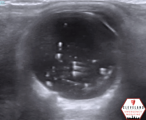
Ophthalmology was consulted for her presenting symptoms. In the meantime, the ultrasound team performed an ocular point-of-care ultrasound (POCUS), primarily to evaluate for retinal or vitreous detachment.


POCUS Findings: There was no evidence of retinal detachment visualized but there are multiple distinct mobile hyperechoic structures within the vitreous body. Findings are somewhat similar to vitreous hemorrhage, but the particles are more discrete and hyperechoic compared to typical findings seen with vitreous hemorrhage/detachment. This is suggestive of asteroid hyalosis.
Case Continued: Ophthalmology performed a comprehensive assessment including a dilated fundoscopic exam. This confirmed asteroid hyalosis as well as mild cataracts and ruled out retinal pathology and vitreous detachment/hemorrhage. Additionally, there was also concern for possible early herpes zoster ophthalmicus. The patient was ultimately deemed stable for outpatient management. She was discharged home with strict return precautions and close follow up was arranged.
Asteroid Hyalosis & Ocular POCUS
Asteroid Hyalosis (AH)
Asteroid hyalosis, named for resembling “stars in the night sky,” is a benign vitreous condition resulting in calcium phospholipid deposits, varying in size, within the posterior chamber [1]. It’s been shown to strongly correlate with increasing age, male sex, and lack of vitreous detachment, but the etiology is not well known. Often asymptomatic, it’s often an incidental finding and typically doesn’t require any intervention [2].
AH vs. Vitreous Hemorrhage (VH)
AH can be easily misinterpreted as vitreous hemorrhage on POCUS as both pathologies produce numerous echogenic opacities and have the classic “washing machine” appearance with extraocular movement. AH tends to have more brighter, more discrete, scintillating particles throughout the vitreous, whereas VH is generally more heterogeneous and layers in the posterior aspect of the vitreous [3-5].

Ocular Ultrasound Technique [4, 6]
Cover the forward-facing closed eye with a thin, clear transparent film (e.g. a Tegaderm). Aattempt to minimize any air bubbles trapped underneath to optimize the image
Apply a copious amount of ultrasound gel over the tegaderm
Place the high-frequency linear probe over the eye with the probe marker pointing to the patient’s right
*Avoid applying pressure excessive pressure by stabilizing your hand on the patient’s face.
Fan the probe throughout the eye and then ask the patient to look left and right (kinetic exam). It’s important to visualize the optic nerve as this can help differentiate pathology
Can also apply this aforementioned technique with the probe in the longitudinal axis. Additionally, compare images with the asymptomatic eye.
When looking within the vitreous body, be sure to increase the gain to a very high level. This helps highlight pathologic findings (see images in the case above), and failing to do this may lead to missed abnormalities
Other pathologies assessed with ocular POCUS include:
Retinal detachment
Time-sensitive and vision-threatening
Appears as a hyperechoic linear membrane attached/tethered to the optic nerve on ocular movement [6-7]

Foreign body
Hyperechoic structure, often seen with reverberation artifact. May produce a “twinkling” artifact with color doppler [6]

Ocular foreign body with reverberation (left) and twinkle artifact (right) [6]
Lens dislocation
Time-sensitive and vision threatening (anterior > posterior). Lens will typically move independently of surrounding structures [7]

Lens dislocation [8]
The Evidence?
Not commonly discussed in the literature; described by a few case reports [4,5].
Take Home Points
Ocular ultrasound can help make timely recognition of multiple emergent ocular conditions and differentiate these from more benign conditions. While AH is benign, it can easily mimic the more potentially serious vitreous hemorrhage on ocular ultrasound. Knowledge of this mimic and ability to recognize the subtle sonographic differences can help differentiate these disease processes, which can influence management and potentially disposition. However, when in doubt, it’s safer to presume VH and manage accordingly until a comprehensive ophthalmology evaluation proves otherwise.
AUTHORED BY: DR. ENIOLA GROS (R1)
FACULTY CO-AUTHOR/EDITOR: LAUREN MCCAFFERTY, MD
References
Khoshnevis M, Rosen S, Sebag J. Asteroid hyalosis: a comprehensive review. Surv Ophthalmol. 2019. 64: 452-462.
Fawzi AA, Vo B, Kriwanek R, Ramkumar HL, Cha C, Carts A, Heckenlively JR, Foos RY, Glasgow BJ. Asteroid hyalosis in an autopsy population: The University of California at Los Angeles (UCLA) experience. Arch Ophthalmol. 2005;123(4):486-90
Kachewar SG, Kulkarni DS. An imaging review of intra-ocular calcifications. J Clin Diagn Res. 2014;8(1):203-5.
Stringer CEA, Ahn JS, Kim DJ. Asteroid Hyalosis: A Mimic of Vitreous Hemorrhage on Point of Care Ultrasound. CJEM. 2017;19(4):317-320.
Lema PC, Mantuani D, Nagdev A, Adhikari S. Asteroid Hyalosis Masquerading as Vitreous Hemorrhage on Point-of-Care Sonography. J Ultrasound Med. 2018; 37(1):281-284.
Jehle Lyon M, von Kuenssberg Jehle D. Ocular. In OJ Ma, JR Mateer, RF Reardon, SA Joing (eds), Ma and Mateer’s Emergency Ultrasound (3rd ed). New York, NY: McGraw-Hill Education. pp 569-586.
Lahham S, Ali Q, Palileo BM, Lee C, Fox JC. Role Of Point Of Care Ultrasound In The Diagnosis Of Retinal Detachment In The Emergency Department. OAEM. 2019; 11:265-270.
Glickman A, Szczucki B, Kalivoda EJ, Furiato A, Cabrera G. Bedside Ocular Ultrasound Diagnosis of a Traumatic Lens Dislocation. Cureus. 2019; 13(4): e14666.