Intern Ultrasound of the Month: Not Just Cellulitis from a Cat Bite… A Case of Extensor Tenosynovitis Diagnosed with POCUS
The Case
30yo M, otherwise healthy, presented to the ED for left hand/wrist pain after sustaining a cat bite/scratch to the dorsum of hand the night before. A piece of the cat’s claw was removed at the time and he washed out the wound with soap and water. He subsequently developed increasing pain, swelling, and limited ROM of the hand and wrist. He was concerned that a claw fragment was still retained. Denied paresthesia, fever, chills, bleeding, purulence, or any other symptoms. Both the patient and the cat are fully vaccinated.
He was well-appearing and in no distress. Vitals WNL. Exam was significant for multiple superficial linear abrasions to the dorsal left hand/forearm, 2 deeper puncture wounds near the base of the 2nd-3rd metacarpals, and mild erythema, edema, tenderness to palpation over the mid-proximal dorsal hand/wrist. He had pain with passive extension of the wrist and MCPs. Wrist/MCP flexion was slightly painful but intact. ROM of digits was fully intact as well. He had no sensory deficits. Digits were well-perfused. There was no fluctuance or drainage from the wounds.
POCUS was performed to primarily to evaluate for abscess and foreign body
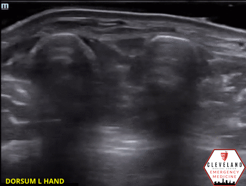
2nd-4th Extensor Tendons with fluid in the tendon sheath
Moving more proximally, the tendons converge at the wrist.

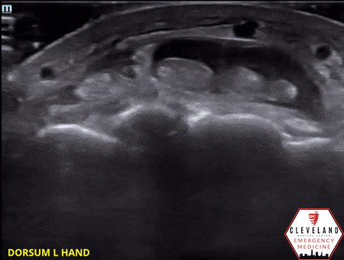
Longitudinal view of the 3rd extensor tendon again demonstrating fluid above and below. 2nd & 4th tendons had similar appearance.
POCUS findings: fluid in the tendon sheath surrounding the extensor tendons along with thickening and mild cobblestoning of the overlying soft tissues. No abscess visualized. Findings suggestive of extensor tenosynovitis.
Case continued: Xray was also obtained and showed diffuse soft tissue swelling but no bony abnormalities. Because of POCUS findings, the patient received IV antibiotics, and hand surgery was consulted. Bedside washout was performed. The patient was admitted for overnight observation and IV antibiotics. He did well without further progression of the infection and was discharged home on augmentin the next day. At an outpatient follow up appointment less than 2 weeks later, symptoms had resolved.
Brief Background on Extensor Tenosynovitis & Cat Bites
The lesser known infectious tenosynovitis
Extensor tenosynovitis
An inflammatory/infectious condition of an extensor tendon +/- tendon sheath
Far less common than flexor tenosynovitis (FTS), & thus, is far less studied & discussed
Similar to FTS, infectious etiology is a result of trauma (i.e. bite wound, puncture wound, laceration) with direct inoculation, hematogenous spread, or contiguous spread, and the most common pathogens are staph aureus & streptococcus [1]
In contrast to FTS, extensor tenosynovitis
Tends to lack classic clinical findings like those of flexor tenosynovitis & is therefore more likely to be missed
May be treated more conservatively, such as with observation & IV antibiotics, rather than surgically
—> this is because the extensor tendons lack a closed retinacular system, so the risk of loculation & increased pressure (potentially resulting in necrosis & rupture) is lower [1-2].
Cat Bites
Higher risk for infection than dog bites due to deeper puncture wound
Polymicrobial — pasturella most common, followed by strep species then staphylococci, Neisseria, & Moraxella [3]
Potential complications: cellulitis, abscess, deep soft tissue infection such as tenosynovitis, osteomyelitis [4]
POCUS Assessment for Tenosynovitis
Technique
High frequency probe
Scan in transverse and longitudinal planes
Identify the tendons — appear as well-organized fibrillar structures with multiple parallel lines in long axis & small circular bundles in short axis. Some tendons, such as those in the hand, are surrounded by a thin synovial sheath which may contain a trace amount of synovial fluid
Fully fan through area of concern
Apply color doppler
A few tips:

Use a water bath to help minimize pain (as this can avoid direct contact with the affected area). This may also help optimize image quality as well.
Compare to the unaffected side, especially if findings are unclear
Beware of anisotropy — a property of tendons in which the echogenicity varies based on angle on insonation. When the probe is perpendicular the to tendon, it will appear hyperechoic but as the probe is fanned, even slightly, the tendon will assume a more hypoechoic appearance. This is a normal finding but can mimic pathologic fluid. [5-6]
POCUS findings of tenosynovitis [7-8]
Tendon and/or tendon sheath thickening & hyperemia with color doppler
Fluid/hypoechoic material within the synovial sheath surrounding the tendon
Thickening &/or cobblestoning of overlying soft tissue


What Does the Evidence Show?
Extensor tenosynovitis is far less studied compared to flexor tenosynovitis in general.
The vast majority of ultrasound evidence looks at the diagnostic accuracy of flexor tenosynovitis with minimal literature, namely case reports, on extensor tenosynovitis [9]
US found to have 94% sensitivity, 74% specificity for early flexor tenosynovitis if either peri-tendinous effusion or thickened synovial sheath were seen. [10]
*This study excluded those patients whose diagnosis was obvious so it supports the utility of US in cases where the clinical picture is less clear.
US had higher sensitivity for detecting inflammation of digits than clinical exam [11]
Take Home Points
Extensor tenosynovitis is a less common and less discussed form of infectious tenosynovitis
While it may have some commonalities with its counterpart, flexor tenosynovitis, extensor tenosynovitis may have a less characteristic clinical presentation & may be managed conservatively
POCUS is a useful tool in diagnosing both forms of infectious tenosynovitis, especially in earlier cases or when clinical picture is not entirely clear.
POST BY: DR. DANI RAO, PGY1
FACULTY EDITING BY: DR. LAUREN MCCAFFERTY
References
Small LN, Ross JJ. Suppurative tenosynovitis and septic bursitis. Infect Dis Clin North Am. 2005;19(4):991–1005–xi.
Newman ED, Harrington TM, Torretti D, Bush DC. Suppurative extensor tenosynovitis caused by Staphylococcus aureus. J Hand Surg Am. 1989;14(5):849-851.
Talan DA, Citron DM, Abrahamian FM, Moran GJ, Goldstein EJ. Bacteriologic analysis of infected dog and cat bites. N Engl J Med. 1999; 340:85–92.
Quinn J. Puncture Wounds and Bites. In: Tintinalli JE, Ma O, Yealy DM, Meckler GD, Stapczynski J, Cline DM, Thomas SH. eds. Tintinalli's Emergency Medicine: A Comprehensive Study Guide, 9e. McGraw-Hill; Accessed April 03, 2021
Lee JC, Healy JC. Normal sonographic anatomy of the wrist and hand. Radiographics. 2005 Nov-Dec;25(6):1577-90
Ma OJ, Mateer JR, Reardon RF, & Joing S. Ma and Mateer's Emergency Ultrasound. New York, NY: McGraw-Hill Education, 2014.
Padrez K, Bress J, Johnson B, Nagdev A. Bedside ultrasound identification of infectious flexor tenosynovitis in the emergency department. West J Emerg Med. 2015;16(2):260-262.
Alcalde M, D'Agostino MA, Bruyn GA, et al. A systematic literature review of US definitions, scoring systems and validity according to the OMERACT filter for tendon lesion in RA and other inflammatory joint diseases. Rheumatology. 2012;51(7):1246-1260.
Frank J A, Lupton J, Hicks B. Point-Of-Care Ultrasound for the Diagnosis of Extensor Tenosynovitis of the Wrist. JETem. 2019; 4(3):V1-4.
Jardin E, Delord M, Aubry S, Loisel F, Obert L. Usefulness of Ultrasound for the Diagnosis of Pyogenic Flexor Tenosynovitis: A Prospective Single-Center Study of 57 Cases. Hand Surg Rehabil. 2018;37(2):95-98.
Hmamouchi I, Bahiri R, Srifi N, Aktaou S, Abouqal R, Hajjaj-Hassouni N. A comparison of ultrasound and clinical examination in the detection of flexor tenosynovitis in early arthritis. BMC Musculoskelet Disord. 2011;12:91.