Intern Ultrasound of the Month: Cellulitis of the Lower Extremity
The Case
35-year-old male with a history of IV drug use who presented to the ED for left lower extremity pain and swelling. He stated that his leg and foot starting to swell the previous day with associated pain with ambulation. He attempted to go to urgent care but due to long wait times, he eventually left prior to being seen. He noted that the swelling had progressed that morning, ultimately convincing him to return to the ED. The patient denied any trauma to his leg and stated that he thought he may have been bitten by a spider, although he did not remember seeing one. He was noted to have superficial abrasions to his left leg, and he stated that he scraped his left shin two days prior while riding a bike. He denied any significant medical history, including HIV or malignancy, and denied any other systemic symptoms. Additionally, he stated that he had been sober and drug-free for several years.
The patient’s vital signs remained stable in the ED, and he did not appear septic. Physical exam revealed left lower extremity edema, erythema, and induration from below the knee through the foot (see photo to the right) with associated tenderness to palpation. However, no crepitus, bleeding, or purulent drainage was noted. As you can see in the photo, the demarcation line was marked by a skin pen for continued monitoring. There were no signs of a bite found. His dorsalis pedis pulse was 1+.
Point-of-care ultrasound (POCUS) was performed to evaluate for abscess and cellulitis given the exam findings and HPI.


POCUS Findings:
There is extensive cobblestoning in the subcutaneous tissues throughout the affected lower extremity without evidence of any focal fluid collections. This signifies cellulitis without signs of abscess or deeper infection.
Cellulitis
Emergency departments see approximately 6 million patients per year for cellulitis or abscess, with the annual incidence of cellulitis ranging from 22 to 50 per 1000 persons (1). In the United States, there are more than 14 million cases every year (2, 3). It is relatively rare that a patient with cellulitis requires hospitalization, happening in less than 10% of cases with an associated mortality rate of 2.5%. The severity and rate of hospitalization increases for patients over the age of 55 (1,4).
Cellulitis is a form of skin and soft tissue infection. The skin is comprised of three layers; the epidermis, dermis, and hypodermis. Cellulitis is a bacterial infection arising from a cutaneous disruption that primarily affects the middle layer of skin, the dermis, and can extend into underlying tissue. This, in turn, causes a robust inflammatory response with neutrophil infiltration and cytokine production (1-2). An abscess, a collection of pus within the dermis or hypodermis, occurs in a similar fashion but can also include phagocytosis, liquefactive necrosis, and edema due to a portal of entry such as a wound or hair follicle. A fibrous capsule then develops, which is often subsequently surrounded by erythema and induration. Purulent cellulitis is when the erythema and induration spread beyond the margins of the abscess (1).
Staphylococcus and Streptococcus are the two most common bacteria that cause cellulitis. Streptococcus species account for greater than 70% of cases, with Group A streptococcus (Strep. pyogenes) being the most common. Staphylococcus Aureus is the next most common, diagnosed in 14%-27% of cellulitis cases. While a common concern in skin infections, methicillin-resistant Staph. aureus (MRSA) only accounts for approximately 4% of nonpurulent cellulitis. On the other hand, S. Aureus is found as the cause for 60-75% of abscesses, with MRSA accounting for up to 70% of these. Polymicrobial abscesses, which include anaerobic microbes, are common in injection drug users (1,5).
Cellulitis is generally a clinical diagnosis, usually presenting with pain, warmth, edema, tenderness, and poorly demarcated erythema. Oftentimes, it can be challenging to differentiate cellulitis from abscess based on physical exam alone, which has treatment implications since an abscess warrants incision and drainage (I&D) rather than antibiotics alone (1-2). Unfortunately, many common diagnostic tests have poor diagnostic utility. For example, white blood cell count, c-reactive protein, erythrocyte sedimentation rate, and procalcitonin are not always elevated and none are specific (2,6). In addition, blood cultures also do not have high yield, as a meta-analysis showed that patients with cellulitis only had a positive blood culture 7.9% of the time (7). Thus, the Infectious Diseases Society of America (IDSA) does not recommend routine blood cultures unless the patient is systemically ill, immunocompromised, or at risk of atypical infections (5). In cases of abscesses, the IDSA does recommend a culture of the purulent fluid - I&D is the primary treatment - though empiric treatment is reasonable in most cases since the majority of these infections are due to MRSA (5). Plain radiographs are neither sensitive nor specific (1)
POCUS for Soft Tissue Infections
In contrast to labs and plain radiographs, POCUS is a highly useful adjunct to the clinical assessment for cases of suspected cellulitis and abscess, particularly in distinguishing between the two. A systematic review and meta-analysis found POCUS to be 98.7% sensitive and 91% specific in differentiating abscess from cellulitis in adults. They also found that POCUS led to a correct change in management in 10.3% of cases with a number needed to treat of 10 (8). By allowing providers to directly visualize whether or not a fluid collection is present, POCUS can indicate the need for I&D or avoid an unnecessary procedure, while also providing guidance for further imaging or involvement of consultants. POCUS may also identify signs of a more serious infection, such as a necrotizing infection or joint involvement, which can help expedite appropriate management (9-10).
Technique & Findings
The high-frequency linear array transducer is typically the most useful to assess for cellulitis and abscess due to the relatively superficial location of the areas affected. In normal skin and soft tissue, the epidermis and dermis sonographically appear as a relatively hyperechoic structure at the top of the screen. Underlying this is the subcutaneous tissue in which adipose has a hypoechoic (dark gray to almost black) appearance with interspersed fine hyperechoic lines of connective tissue. Deep to this are fascial planes, appearing as distinct hyperechoic membranes, and muscle tissue which has a linear fibrous appearance in long axis. Finally, just below the muscle layer is the highly hyperechoic cortical bone with posterior shadowing due to its highly reflective surface (11-12). Understanding this sonoanatomy is important for soft tissue POCUS evaluations for cellulitis and abscesses; for both, the area of focus is in the more superficial layers.

Figure 1. Sonoanatomy - Layers of healthy skin and soft tissue in long (left) and short (right) axis. Image from Burgin 2020 (11)
Scanning for cellulitis is best accomplished by scanning in two planes throughout the affected area, in addition to areas both proximal and distal to this. Cellulitis findings on ultrasound progress through stages based on severity. In early cellulitis, the subcutaneous tissue appears thickened and has increased echogenicity with blurring or delineation of the typical sharp edges of soft tissue planes, see Figure 2. As it progresses, there will be more fat stranding and fluid accumulating in the subcutaneous tissue. Mature cellulitis appears as fluid in the interstitial space between the more hyperechoic fat lobules, described as “cobblestoning” due to its similarity to a cobblestone street (11-12); see Figure 2. It is also important to note that cobblestoning is not specific and can be seen in cases of lymphedema or volume overload (11).

Figure 2. Early cellulitis

Figure 3. Cobblestoning (ultrasound image from our patient), as seen in more mature stages of cellulitis, has the appearance of cobblestones on a street
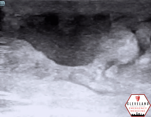
Figure 4. Abscess with swirling of debris seen with compression
As mentioned previously, discerning an abscess from cellulitis is a key role for POCUS. An abscess appears as a localized collection of fluid typically within the loose connective layer of subcutaneous adipose. It is often largely anechoic or hypoechoic but may contain echogenic debris and often has posterior acoustic enhancement (11-12). It is important to assess the extent of a distinct fluid collection by fanning through the entire structure in two planes. To help verify the presence of an abscess using POCUS, apply intermittent pressure over the area of concern and assess for compressibility. Being a pocket of fluid, an abscess will “squish”, whereas non-localized soft tissue edema or solid structures will not (11). In addition, purulent material can be visualized on ultrasound as echogenic debris that swirls with compression or the presence of loculations, which can help differentiate an abscess from other mimics, such as a simple cyst. It is important to apply color doppler over a fluid collection to ensure it is not a vascularized structure, such as a pseudoaneurysm, and to identify surrounding vasculature and other structures, especially when considering I&D. For both cellulitis and abscess, it is imperative to take into account the clinical context and consider potential mimics on ultrasound, such as non-infectious edema, lymph nodes, vasculature, or even herniated bowel (11-12).
Necrotizing fasciitis is a rapidly progressing, life-threatening soft tissue infection that needs timely surgical intervention. In early stages, the findings can mimic those of cellulitis. However, as the infection progresses, fluid along the fascial plane will develop, see Figure 5. A late, but pathognomonic, finding is the presence of air in the subcutaneous tissues. Sonographically this appears as hyperechoic foci with “dirty shadowing” (10).

Figure 5. Necrotizing fasciitis - fluid along the fascial plane

Figure 6. Necrotizing fasciitis with subcutaneous air/dirty shadowing, a late finding
Case Conclusion:
An x-ray was performed and was negative for gas, fracture, or foreign body. Lab work revealed a leukocytosis of 13 with a neutrophil predominance and normal lactate. POCUS findings were used in conjunction with the other clinical findings to support a diagnosis of cellulitis. The patient was given broad spectrum antibiotics along with IV fluids. He was then admitted for observation and continued treatment with IV antibiotics. After twenty-four hours, his vitals remained stable, pain was well-controlled with ibuprofen, and there was regression of the infection from the delineated border. Having met those criteria, he was discharged on keflex and ibuprofen with a follow-up appointment with his PCP in one week.
Take Home Message
Cellulitis is a clinical diagnosis but POCUS is a great adjunct to your physical exam, especially when the clinical picture is not obvious. POCUS is especially useful in differentiating cellulitis from abscess, which can guide treatment and indicate need for additional imaging or surgical consultation.
POST BY: DAVID WILLIAMS, DO, PGY1
FACULTY CO-AUTHOR/EDITOR: LAUREN MCCAFFERTY, MD
References
Long, B., Gottleib, M. Diagnosis and Management of Cellulitis and Abscess in the Emergency Department Setting: An Evidence-Based Review. J Emerg Med. 2022; 62(1): 16-27.
Raff AB, Kroshinsky D. Cellulitis: a review. JAMA. 2016; 316:325–37.
Brown BD, Hood Watson KL. Cellulitis. [Updated 2023 Aug 7]. In: StatPearls [Internet]. Treasure Island (FL): StatPearls Publishing; 2023. Available from: https://www.ncbi.nlm.nih.gov/books/NBK549770/
McNamara DR, Tleyjeh IM, Berbari EF, et al. Incidence of lower-extremity cellulitis: a population-based study in Olmsted county, Minnesota. Mayo Clin Proc. 2007; 82:817–21.
Stevens DL, Bisno AL, Chambers HF, et al. Practice guidelines for the diagnosis and management of skin and soft tissue infections: 2014 update by the Infectious Diseases Society of America. Clin Infect Dis. 2014;59(2):e10–52.
Lazzarini L, Conti E, Tositti G, de Lalla F. Erysipelas and cellulitis: clinical and microbiological spectrum in an Italian tertiary care hospital. J Infect. 2005; 51:383–9.
Gunderson CG, Martinello RA. A systematic review of bacteremias in cellulitis and erysipelas. J Infect. 2012; 64(2):148-155.
Gottlieb M, Avila J, Chottiner M, Peksa GD. Point-of-care ultrasonography for the diagnosis of skin and soft tissue abscesses: a systematic review and meta-analysis. Ann Emerg Med. 2020;76(1):67– 77.
Adhikari S, Blaivas M. Utility of bedside sonography to distinguish soft tissue abnormalities from joint effusions in the emergency department. J Ultrasound Med. 2010; 29:519–526.
Gan RK, Sanchez Martinez A, Abu Hasan MA, Castro Delgado R, Arcos González P. Point-of-care ultrasonography in diagnosing necrotizing fasciitis-a literature review. J Ultrasound. 2023; 26(2):343-353
Burgin C, Morrow D. Utility of POCUS in skin and soft tissue infection. J Urgent Care Med. June 2020; 17-21.
Euerle B. “Abscess Evaluation”. American College of Emergency Physicians Sonoguide. Aug 2020. Available from: https://www.acep.org/sonoguide/procedures/abscess-evaluation