Intern Ultrasound of the Month: Small (& Large) Bowel Obstruction
THE CASE
75yo F presented to the ED for abdominal pain. Her past medical history was significant for recently diagnosed adenocarcinoma of the colon with liver metastases. A CT scan obtained during her recent hospitalization showed a partial small bowel obstruction, which was managed conservatively. The patient stated that shortly after she was discharged, she began to experience generalized abdominal pain, described as waxing and waning, 5/10 in severity. She also reported poor appetite and lack of bowel movements with minimal flatus during the preceding 4-5 days. There was no other associated symptomatology.
On arrival to the ED, her vitals were remarkable for tachycardia in the 120s. Physical exam revealed a non-peritonitic abdomen that was distended and tender to palpation in all four quadrants.
POCUS was utilized to assess for signs of bowel obstruction as well as free fluid.

Dilated, echogenic fluid-filled loops of small bowel with to-and-fro movement of contents. The bowel wall also appears thickened.

Swirling of bowel contents

Bowel diameter was increased at ~4.3 cm

Ascending large bowel (noted by the haustra) is also dilated, measuring over 7cm in the RLQ, with dense intraluminal contents with poor peristalsis

POCUS findings
In the first row of images, you can see well-defined, dilated loops of bowel (with diameter measuring over 4cm), to-and-fro or “swirling” movements of intraluminal bowel contents, and bowel wall edema. The ascending colon (in the 2nd row of images) also appears dilated, measuring over 7cm. Not shown here but the majority of the transverse and descending colon did not appear dilated, suggesting a transition point though this was not visualized.
Findings are concerning for small (and large) bowel obstruction. Of note, there was no evidence of free fluid.
Case continued
Basic labs were obtained and were largely within normal limits. Abdominal xray found no evidence of pneumoperitoneum but showed dilation of the ascending colon. CT abdomen/pelvis confirmed the diagnosis of high-grade small and large bowel obstruction. She was kept NPO and an NG tube was placed for decompression, which helped alleviate her symptoms. Colorectal surgery was consulted and she was admitted to the hospital for further management. She underwent successful hemicolectomy, ileocolic anastomosis, and diverting loop ileostomy. Her post-op course was uncomplicated, and she was discharged home a few days later.
Small Bowel Obstruction, A Brief Review
Epidemiology
Small bowel obstruction (SBO) is frequently encountered by emergency physicians. In the United States, approximately 300,000 adults are hospitalized annually for SBO [1-2].
SBO accounts for approximately 2% of emergency department visits, 15% of hospital admissions, and 20% of emergent surgical operations for abdominal pain [2-4]
Etiology
Most common underlying cause is adhesions from a prior abdominal surgery, accounting for 60-75% of cases. This is followed by colonic neoplasm, hernia, IBD, volvulus [5-6]
Clinical Presentation
Most common symptom is loss of passage of stool +/- flatus, and the most common sign is abdominal distention [6]. Other common signs/symptoms include abdominal pain, nausea, vomiting, peritoneal signs, abdominal distention, tachycardia, and leukocytosis.
Signs and symptoms can vary based on degree of obstruction, location of the obstruction (proximal vs distal), and accompanying pathology/comorbid conditions [5-6].
Diagnostic Imaging
CT is considered the gold standard but incurs cost, radiation exposure, and time and is not always readily available. Xrays may be a useful initial imaging modality but generally poor diagnostic accuracy [2]
Ultrasound has shown promising utility
POCUS for Bowel Obstruction
Indications
Any patient with the above signs/symptoms, but ultrasound is particularly well-suited for patients in whom clinical suspicion for SBO is high due to clinical presentation and risk factors.
Focused Clinical Question(s)
Is there evidence of obstruction? see below
Can also assess for possible location and/or causes of the obstruction
Technique
Probe selection: low frequency curvilinear probe is preferred
Patient positioning: supine position is usually best
Use a systematic approach, such as “mowing the lawn,” whereby the probe is placed in the transverse orientation in the lateral edge of one quadrant and you slide the probe up and down the abdomen from the level of the epigastrium to the pelvis, moving from one side of the abdomen to the other
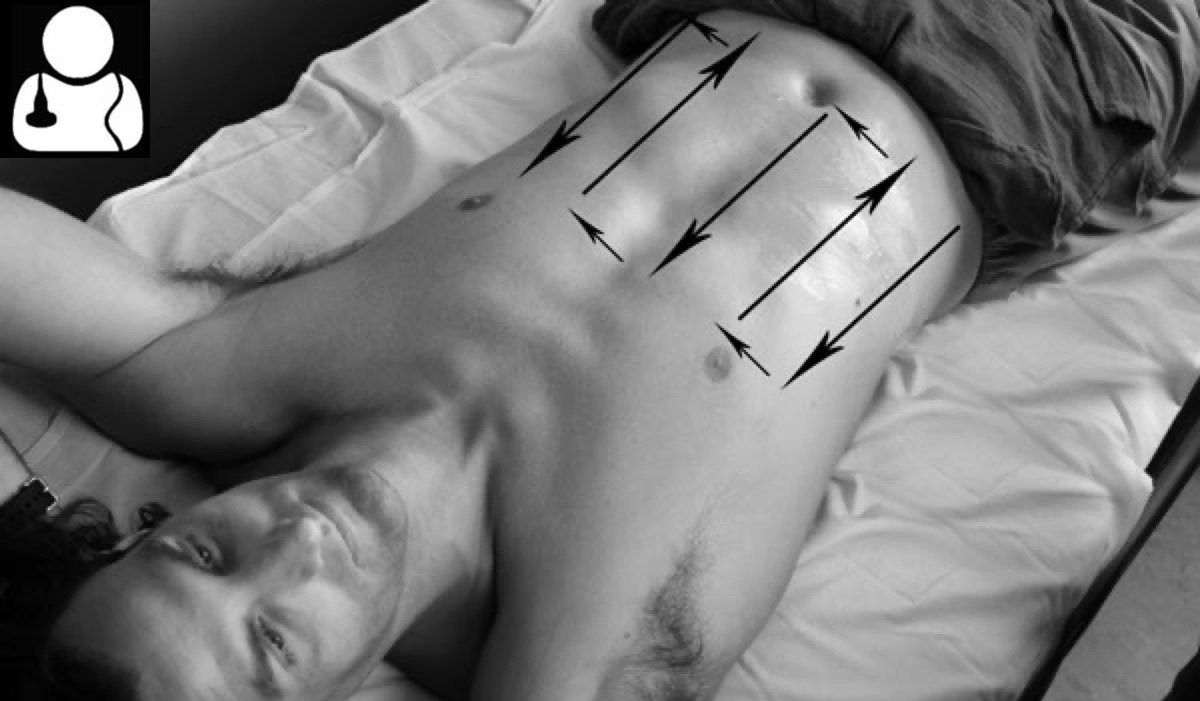
“Mowing the lawn” approach. Image from: https://www.coreultrasound.com/uotw-20-answer/
Ultrasound findings suggestive of SBO [7-10]
Small bowel dilation >2.5 cm, measured outer wall to outer wall)
Abnormal peristalsis — “to-and-fro” movement/swirling of intraluminal contents or lack of peristalsis)
Bowel wall edema (could indicate ischemia) +/- prominent plicae circulares (“keyboard sign”)
Transition point — dilated loops of bowel next to decompressed (poorly visualized) bowel
Intraperitoneal free fluid — suggests high grade obstruction or possible perforation [11]
Most sensitive finding = dilated bowel >2.5 cm; most specific findings = transition point & free fluid [7]

Free fluid between loops of bowel - “Tanga Sign” - associated with high-grade obstruction
**Large Bowel Obstruction will show dilated colon (noted by haustra) along the periphery of the abdomen measuring >5 cm (ascending colon) [10].
Limitations/Pitfalls
Operator dependence
Body habitus
Don't confuse small and large bowel. Large bowel has characteristic haustra (echogenic curvilinear arcs of colonic wall) which give it a segmental appearance
Bowel gas or air from air-fluid levels or perforation may preclude good visualization of bowel. If this is the case, consider a more lateral or inferior approach since air tends to layer in the least dependent areas (anteriorly if supine or superiorly if upright)
Utility of Ultrasound for SBO
Ultrasound has shown good diagnostic accuracy for SBO and significantly outperforms x-ray [2]. A meta-analysis by Gottlieb et al found US to have a sensitivity of 92.4% and specificity of 96.6% specific [12]
When utilized by ED physicians to assess for SBO, POCUS has demonstrated sensitivity of 88% and specificity of 54%. However, diagnostic parameters are significantly higher when the ultrasonographer is fellowship trained [7], suggesting more training and familiarity with this assessment can significantly improve accuracy. This is supported by another study of emergency physicians that required more focused SBO experience prior to participating in the study and had higher diagnostic accuracy [13].
POCUS incurs minimal to no risk (such as from ionizing radiation, IV contrast) to the patient and can be done quickly at the bedside. While it doesn't necessarily replace CT, it can give you a diagnosis within minutes rather than hours and can allow the provider to initiate targeted management and reach out to necessary consultants earlier.
Take Home Points
SBO is a frequently encountered diagnosis in the ED and is a common reason that patients with abdominal pain are hospitalized and/or undergo surgery
Consider SBO in any patient with abdominal pain/distention, nausea, vomiting, obstipation, etc. especially if he/she has high risk features
Sonographic findings suggestive of SBO: more clearly visualized bowel anatomy, dilated fluid-filled small bowel measuring >2.5 cm in diameter, abnormal persistalsis or “to-and-fro” movements of intraluminal contents, bowel wall edema, transition point. Intraperitoneal fluid suggests high-grade obstruction
Ultrasound is quite sensitive but less specific for SBO when performed by ED physicians. Diagnostic accuracy improve significantly with more ultrasound training
CT remains the gold standard in diagnosing SBO, however, POCUS can be used as a quick bedside tool to help expedite diagnosis and management
AUTHORED BY: DR. WILL HEERSINK (PGY1)
FACULTY CO-AUTHOR/EDITOR: LAUREN MCCAFFERTY, MD
REFERENCES
Hastings RS, Powers RD. Abdominal pain in the ED: a35 year retrospective. Am J Emerg Med. 2011;29:711–6.
Taylor MR, Lalani N: Adult small bowel obstruction. Acad Emerg Med. 2013, 20:528-44
Cappell MS, Batke M. Mechanical obstruction of the small bowel and colon. Med Clin North Am. 2008; 92(3):575-97, viii.
Gore RM, Silvers RI, Thakrar KH, Wenzke DR, Mehta UK, Newmark GM, Berlin JW. Bowel Obstruction. Radiol Clin North Am. 2015;53(6):1225-40.
Jackson P, Vigiola Cruz M. Intestinal obstruction: evaluation and management. Am Fam Physician. 2018; 98:362-367.
Markogiannakis H, Messaris E, Dardamanis D, et al. Acute mechanical bowel obstruction: clinical presentation, etiology, management and outcome. World J Gastroenterol. 2007;13(3):432-437.
Becker BA, Lahham S, Gonzales MA, Nomura JT, Bui MK, Truong TA, et al. A Prospective, Multicenter Evaluation of Point-of-care Ultrasound for Small-bowel Obstruction in the Emergency Department. Acad Emerg Med. 2019 Aug;26(8):921-930.
Pourmand A, Dimbil U, Drake A, Shokoohi H. The Accuracy of Point-of-Care Ultrasound in Detecting Small Bowel Obstruction in Emergency Department. Emerg Med Int. 2018;2018:3684081.
Abu-Zidan, F.M., Cevik, A.A. Diagnostic point-of-care ultrasound (POCUS) for gastrointestinal pathology: state of the art from basics to advanced. World J Emerg Surg. 2018; 13, 47.
Ogata M. General Surgery Applications. In OJ Ma, JR Mateer, RF Reardon, SA Joing (eds), Ma and Mateer’s Emergency Ultrasound (3rd ed). New York, NY: McGraw-Hill Education. pp 273-317.
Grassi R, Romano S, D’amario F, et al. The relevance of free fluid between intestinal loops detected by sonography in the clinical assessment of small bowel obstruction in adults. Eur J Radiol. 2004;50(1):5-14
Gottlieb M, Peksa GD, Pandurangadu AV, Nakitende D, 2, Takhar S, Seethala RR. Utilization of ultrasound for the evaluation of small bowel obstruction: A systematic review and meta-analysis. Am J Emerg Med. 2018;36(2):234-242.
Jang T. B., Schindler D., Kaji A. H. Bedside ultrasonography for the detection of small bowel obstruction in the emergency department. Emerg Med J. 2011;28(8):676–678