Intern Ultrasound of the Month: Retinal Detachment
The Case
55-year-old relatively healthy male presented to the emergency department for painless vision changes in the left eye. He stated he first noticed black spots in his left eye about 10 days prior arrival and then a few days later started having decreased visual acuity in the inferior visual field; described as a “curtain coming down” his eye. He denied any pain with extraocular movements, fever, chills, headache, dizziness, nausea, vomiting, URI symptoms, focal neurologic deficits, preceding illness or recent trauma. ROS was otherwise negative.
Vitals were stable on arrival. Physical exam was relatively unremarkable. His pupils were equal and reactive and extraocular movements intact. Acuity: OS 20/25 (OD 20/20). Intraocular pressure was normal. Ocular exam was negative for the following: conjunctival injection, drainage, direct or consensual photophobia, afferent pupillary defect, visualized foreign body, proptosis, periorbital edema or signs of cellulitis. Neuro exam was intact as well.
Ocular POCUS was performed to evaluate for vitreous pathology (retinal or vitreous detachment, vitreous hemorrhage) and revealed the following:


POCUS findings: hyperechoic linear membrane within the vitreous body attached posteriorly near the optic nerve on the medial side of the globe; moves with kinetic exam but remains tethered (is not free floating) —> suggestive of (medial) retinal detachment
Case continued: Ophthalmology was consulted. They performed a comprehensive exam and further supported the diagnosis. The patient was urgently taken to the OR and medial retinal detachment was confirmed.
Retinal Detachment
Brief Overview[1]
What Is It? Separation of the inner light sensing layer of the retina from the pigmented layer. Most commonly due to retinal tear, resulting in fluid accumulating and causing the separation.
Epidemiology —Incidence ~1 in 10,000. Affects men more than women & older age.
Clinical Presentation — painless monocular vision changes -- flashes, floaters most common. Can also cause a “veil” or “curtain” over visual field, decreased visual acuity especially peripherally.
Risk factors — history of retinal detachment, eye surgery, myopia, trauma, posterior vitreous detachment, etc.
Diagnosis — comprehensive ophthalmologic/ fundoscopic exam. Ocular ultrasound has also been used as good adjunct for decades (referred to as “B scan” by ophthalmologists)
Prognosis — potentially vision-threatening. Why it’s considered an ocular emergency, especially if the macula is still attached.
Treatment — surgery (vitrectomy) most common
Ocular Ultrasound [2-4]
Indications
Vision changes
Eye pain
Foreign body
Eye trauma
**Contraindication: concern for globe rupture
General Technique
Linear probe (high frequency), probe marker to patient right
Cover eye with tegaderm (minimize air bubbles)
Apply a lot of gel
Stabilize your hand on pt’s face to control amount of pressure applied
Identify structures of eye
Ask the patient to look side to side (dynamic or kinetic exam)
Look for abnormal findings discussed below
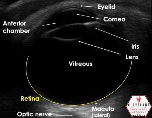
Retinal Detachment
Hyperechoic membrane within the vitreous that’s tethered to the optic nerve posteriorly
Mobile/appears like “the wave” w/ dynamic exam; less free-floating than vitreous detachment
Shouldn’t cross midline (attachment points should be on the same side of globe)
“Mac-on” vs “Mac off” retinal detachment — if the retina still attached to the macula (“Mac on”; the attachment point will be more lateral to the optic nerve), central vision is still preserved so emergent repair is critical in order to prevent progression to macula detachment (Mac off)
Compare to the less-emergent, often age-related:
Vitreous Detachment
Wavy, hyperechoic membrane within the vitreous that’s NOT attached to the optic nerve; can cross over midline. Often thinner than RD.
May or may not have a more swirling appearance like VH

Vitreous Hemorrhage
Echogenic material within the vitreous swirls with kinetic exam (has a washing machine appearance). Degree of echogenicity is variable.
May be present in isolation or concurrently with RD or VD


A Few Scanning Pearls
Increase the gain to help visualize structures within the vitreous. Could miss abnormal findings otherwise
Perform a dynamic/kinetic exam. This is key for picking up intravitreous abnormalities (i.e. retinal or vitreous detachments, hemorrhage, etc) and distinguishing between them
Visualize the optic nerve to differentiate retinal vs vitreous detachment
*Any membranous attachment near the optic nerve should raise concern for retinal detachment and prompt ophtho evaluation.
What Does the Evidence Show?
Emergency physician-performed POCUS for detecting retinal detachment has been shown to have very high sensitivity (96%-97) with good, but slightly lower, specificity 88-92% based on prospective studies [5-6]
In a recent systemic review and meta-analysis, emergency physicians demonstrated a slightly higher specificity of 96% (with relatively similar sensitivity) for diagnosing retinal detachment with POCUS [7]
Why Does This matter?
Retinal detachment is a vision-threatening and, therefore, time-sensitive diagnosis. While POCUS may not replace a thorough ophthalmology assessment, it can serve as a good adjunct and help expedite diagnosis and appropriate management. This is especially helpful when ophthalmology is not always readily available.
Ocular US is also deemed a core application for emergency physicians [8]
POST BY: DR. CONNOR PARSELL, PGY1
FACULTY EDITING BY: DR. LAUREN MCCAFFERTY
References
1. Steel D. Retinal detachment. BMJ Clin Evid. 2013;1-32.
2. De La Hoz Polo M, Torramilans Lluís A, Pozuelo Segura O, Anguera Bosque A, Esmerado Appiani C, Caminal Mitjana JM. Ocular ultrasonography focused on the posterior eye segment: what radiologists should know. Insights Imaging. 2016;7(3):351-364.
3. Ma OJ, Mateer JR, Reardon RF, & Joing S. (2014). Ma and Mateer's Emergency Ultrasound. New York, NY: McGraw-Hill Education
4. Nagdev, A. Ocular Ultrasound: Retinal Detachment and Posterior Vitreous Detachment. Academic Life in Emergency Medicine. http://www.aliem.com/ocular-ultrasound-retinal-detachment-posterior-vitreous-detachment/ Published March 11, 2014. Accessed December 9, 2020.
5. Lahham S, Shniter I, Thompson M, et al. Point-of-Care Ultrasonography in the Diagnosis of Retinal Detachment, Vitreous Hemorrhage, and Vitreous Detachment in the Emergency Department. JAMA Netw open. 2019;2(4):e192162.
6. Shinar Z, Chan L, Orlinsky M. Use of Ocular Ultrasound for the Evaluation of Retinal Detachment. J Emerg Med. 2011;40(1):53–57.
7. Gottlieb M, Holladay D, Peksa GD. Point-of-Care Ocular Ultrasound for the Diagnosis of Retinal Detachment: A Systematic Review and Meta-Analysis. Acad Emerg Med. 2019;26(8):931-939.
8. American College of Emergency Physicians. Ultrasound Guidelines: Emergency, Point-of-Care and Clinical Ultra- sound Guidelines in Medicine. Ann Emerg Med 2017;69: e27–54