Intern Ultrasound of the Month: Inguinal Hernia
The Case
50yo M with PMH of hypertension, CAD, prior GSW to abdomen s/p ex-lap & pelvic fixation, chronic bilateral inguinal hernias (typically asymptomatic) who presented to the emergency department for 3 days of severe intermittent pain in his right groin. Reported that it felt like his hernia “popped out and won’t go back in.” Also reported dry cough which easily exacerbated his pain. ROS otherwise only positive for nausea and a few episodes of vomiting. Denied fever, chills, vomiting, diarrhea, constipation or obstipation, urinary symptoms, scrotal pain/swelling.
On exam, his vitals were stable, he was sitting up and leaning forward in bed, appeared very uncomfortable. Abdomen was soft, nontender, not peritonitic. A firm, palpable mass, tender to palpation, was noted in the right inguinal region. Not initially reducible with applied pressure. No overlying skin changes. Exam was otherwise reassuring.
Labs and CT abdomen/pelvis were ordered, and the patient received analgesics.
In the meantime, point-of-care ultrasound of the right inguinal mass showed the following:

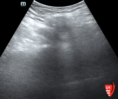
POCUS findings: Peritoneal defect with protruding sac containing loop of bowel and a small amount of free fluid; there’s some peristalsis of the herniated bowel. The image on the right shows the herniated sac reducing with applied pressure. Not shown here but a few dilated loops of bowel in the abdomen were also seen.
CT findings were consistent with ultrasound — bowel-containing right inguinal hernia and signs of focal obstruction with proximally dilated bowel. Also, a small left inguinal hernia containing part of the bladder was seen.

Acute care surgery was consulted and recommended a PO challenge, which the patient easily tolerated. Surgery ultimately opted for elective, rather than urgent, repair (of bilateral hernias), as the patient was not demonstrating significant clinical signs of obstruction.
Hernias

https://www.mountnittany.org/articles/healthsheets/7122

https://www.2minutemedicine.com/patient-basics-hernia/
What is it? — protrusion of fatty tissue and/or bowel through in a weak spot in the abdominal wall muscles; for inguinal hernias, this is generally due to a defect in the inguinal canal wall
Risk factors include: male gender, older age, caucasian, history of hernia, family history of hernia, obesity, smoking, constipation or increased intraabdominal pressure, pre-existing weakness in the abdominal wall
Symptoms commonly include a bulge, pain, swelling
Vomiting, fever, systemic illness may develop in more severe cases
Location/Type of Hernia
See image above
Indirect vs direct inguinal hernia determined by its relationship to the inferior epigastric vessels
Complications
Incarceration: herniated bowel gets trapped, leading to bowel obstruction
Strangulation: blood supply gets cut off resulting in ischemia/necrosis of affected bowel
Diagnosis: most commonly by physical exam; dynamic exam such as Valsalva or coughing, especially with patient standing can aid in evaluation
CT, MRI, ultrasound may be needed if exam is nondiagnostic and if there is concern for complication
Treatment:
Urgent surgical repair indicated if complications are present, particularly strangulation
Elective repair an option if no complications & reduction is possible
Hernia Assessment Using Point-of-Care Ultrasound
Technique
High frequency probe (linear is best, curvilinear if patient has a larger body habitus)
Start by placing the probe in short axis (probe marker to patient right) over the point of maximal pain/swelling and scan the surrounding area
Augment exam by having patient stand, cough, Valsalva, etc.

https://www.coreultrasound.com/uotw-44/
Evaluate for the following:
Peritoneal defect
Hernia contents - bowel vs fatty tissue
Presence or absence of peristalsis in herniated bowel loop
Wall thickness of herniated bowel loop (>3mm is abnormal)
Free fluid in and/or around the hernia sac
Signs of (proximal) bowel obstruction in abdomen
Fluid-filled bowel, diameter >2.5 cm
Decreased or absent peristalsis
Prominent plicae circulares extending from bowel wall - "keyboard sign"
Thickened bowel wall
Free fluid between loops of bowel - "tanga sign"
Distally collapsed bowel suggesting transition point
What does the evidence show?
Meta analysis of ultrasound’s diagnostic accuracy for inguinal hernias (Robinson et al, 2013)
Overall sensitivity 96.6 %, specificity 84.8%, positive predictive value 92.6%
In diagnosing occult hernias, ultrasound demonstrated higher sensitivity/specificity than CT (Robinson et al, 2012)
US sensitivity 86%, specificity 77% vs CT sensitivity 80%, specificity 65%
Findings most predictive of incarceration: free fluid in the hernia sac, increased bowel wall thickness & fluid-containing bowel within the hernia sac (Rettenbacher et al, 2001)
Findings suggestive of strangulation not well studied but thought to include lack of peristalsis in the incarcerated segment, thickened bowel wall (> 3mm), free fluid around the segment. Lack of blood flow on color doppler not very reliable.
Take Home Message
POCUS is useful in the diagnosis (& reduction) of abdominal wall hernias, especially for the more subtle ones; it’s a good adjunct for physical exam/clinical findings and can expedite management & disposition
POST BY: DR. LEONARD KELLER, PGY1
FACULTY EDITING BY: DR. LAUREN MCCAFFERTY
References
2. Noble V, Nelson B. Manual of Emergency Medicine and Critical Care Ultrasound, 2nd ed
4. Fitzgibbons RJ, Forse RA. Groin hernias in adults. N Engl J Med. 2014;372(8):756-763.
5. Robinson A, Light D, Nice C. Meta-analysis of sonography in the diagnosis of inguinal hernias. J Ultrasound Med. 2013;32(2):339-346.
6. Robinson A, Light D, Kasim A, Nice C. A systematic review and meta-analysis of the role of radiology in the diagnosis of occult inguinal hernia. Surg Endosc. 2013;27(1):11-18.
7.. Rettenbacher T, Hollerweger A, Macheiner P, et al. Abdominal wall hernias: Cross-sectional imaging signs of incarceration determined with sonography. Am J Roentgenol. 2001;177(5):1061-1066.