Intern Ultrasound of the Month: Achilles Tendon Rupture
The Case
50 yo M presented to the ED for severe left ankle/calf pain that started while playing basketball. He stated he heard a “loud pop” and simultaneously felt significant pain along the posterior lower leg. Since then he had difficulty moving his ankle or bearing weight due to pain.
On arrival to the ED, his vitals were normal. His physical exam was notable for severe tenderness to palpation of the left calf down to the calcaneous. He was unable to dorsiflex or plantarflex and ROM of the left ankle was limited due to pain. He had no sensory deficits and no overlying skin changes or open wounds.


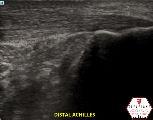
POCUS findings: achilles tendon is visualized here in its longitudinal axis. About 6cm from the calcaneal attachment, there is enlargement of the tendon with disruption of the tendon fibers with retracted edges and multiple hypoechoic areas within the tendon. The proximal tendon (right image) is intact.
Findings are consistent with tendon rupture.
Case continued: Xrays of his ankle were unremarkable. Ortho was consulted and helped facilitate close outpatient follow up for definitive management. The patient was placed in a posterior short leg splint and advised to maintain non-weight-bearing status. An outpatient MRI was performed a few days later and confirmed the diagnosis of full thickness tendon tear. He underwent surgical repair later that week.

Achilles Tendon Rupture
Epidemiology
Incidence of Achilles tendon rupture in the United States is 2.1 per 100,000 person-years and is on the rise
Higher incidence in men
Acute rupture most common between ages 20-40 years old [1]
Vast majority occur during recreational sports
Risk factors: pre-existing achilles tendon pathology, degenerative changes, rheumatologic disease, steroid or fluoroquinolone use [1-2]
Mechanism/Pathophys
Often the result of sudden unexpected dorsiflexion or forced dorsiflexion of planter flexed foot
Most Achilles tendon ruptures occur 2-6 cm above the heel. [2-3]

Diagnosis
Generally a clinical diagnosis but missed in over 20% of cases, possibly because of pain limiting the exam [4]
History
Sudden pop or snap
Sudden onset of severe pain at Achilles tendon region
Exam findings:
Limited planter flexion/ increased passive dorsal flexion
Palpable gap in Achilles tendon
Possible ecchymosis and calf atrophy
Thompson Test: Squeeze affected calf
Positive = absent or reduced planter flexion
Other imaging to consider:
Xrays to rule out other pathology
Ultrasound and MRI can help in confirming and better assessing severity (partial vs full tear)
If not promptly diagnosed, it can result in poor operative outcomes and prolonged disability [2,4]
Management
Conservative management
Posterior short leg splint in planter flexion
Goal is to bring the damaged ends of tendon together
Transition to CAM/walking boot
Operative
Treatment of choice for young active individuals, especially elite athletes
Reduces relatively rare risk of re-rupture but increased rates of infection [5]
Overall debate about which method is best. Regardless, early rehab is key [4,6]
POCUS Assessment for Achilles Tendon Tear
![Adhikari et al. 2012 [7]](https://images.squarespace-cdn.com/content/v1/59b8d169cd39c306cd5e5074/1624465921695-3B3ET1F5VRRMD1PT609N/gr3.jpg)
Adhikari et al. 2012 [7]
Technique
High frequency linear probe is best
Optimize patient position to best expose posterior aspect of the leg (i.e. place in prone position, have foot hanging off bed)
Obtain longitudinal and transverse views
Identify tendon by its classic appearance — linear fibrillar appearance in long axis and oval appearance in short axis
Scan length of the tendon from calcaneous to calf muscle in both planes


Look for:
Disruption in the tendon fibers — may appear thickened, wavy
Secondary signs of injury such as fluid (usually anechoic) representing hematoma, posterior acoustic shadowing at the margins of the rupture, etc. [4, 7]
**Thickened tendon, posterior shadowing, tendon retraction correlate with full thickness tears [4]

A Few Pearls & Pitfalls
Compare affected side to unaffected side
Augment exam with dynamic assessment (slight dorsiflexion & plantar flexion) — movement of the tendon ends away from each other suggests full thickness tear [7]
Beware of anisotropy — a common property of tendons whereby the echogenicity of the tendon changes based on the angle of the probe. Can mimic fluid.
What Does the Evidence Show?
Ultrasound has demonstrated sensitivity of 96-100% and specificity of 83-100% for detecting achilles tendon rupture [4, 8]
Take Home Points
Prompt diagnosis of Achilles tendon rupture is key!
Ultrasound is a great diagnostic tool, especially when the clinical assessment is equivocal — it’s quick, safe, easily accessible, cost-effective, fairly good diagnostic accuracy
Obtain sagittal and transverse views. Scan length of tendon
Look for disruption in normal tendon architecture & adjacent fluid
AUTHORED BY: DR. WESLEY GALLAHER, PGY1
FACULTY CO-AUTHOR/EDITOR: LAUREN MCCAFFERTY, MD
References
Lemme NJ, Li NY, Defroda SF, Kleiner J, Owens BD. Epidemiology of Achilles Tendon Ruptures in the United States Athletic and Nonathletic Injuries From 2012 to 2016. 2016:1-7.
Rose N, Green T (2017). Ankle and Foot. In Rosen’s Emergency Medicine: Concepts and Clinical Practice (9th Ed, p.634-658).Elsevier.
Hess GW. Achilles tendon rupture: a review of etiology, population,anatomy, risk factors, and injury prevention. Foot Ankle Spec. 2010;3(1):29-32.
Hartgerink P, Fessell DP, Jacobson JA, van Holsbeeck MT. Full versus partial-thickness Achilles tendon tears: sonographic accuracy and characterization in 26 cases with surgical correlation. Radiology. 2001;220:406-12.
Ochen Y, Beks R B, van Heijl M, Hietbrink F, Leenen L P H, van der Velde D et al. Operative treatment versus nonoperative treatment of Achilles tendon ruptures: systematic review and meta-analysis. BMJ. 2019; 364:k5120
Buddecke D. Acute Achilles Tendon Ruptures. Clin Podiatry Med Surg. 2021; 38:201-226.
Adhikari S, Marx J, Crum T. Point-of-care ultrasound diagnosis of acute Achilles tendon rupture in the ED. Am J Emerg Med; 2012; 30(4): 634.e 3-4.
Paavola M, Paakkala T, Kannus P, Jarvinen M. Ultrasonography in the differential diagnosis of Achilles tendon injuries and related disorders. A comparison between pre-operative ultrasonography and surgical findings. Acta Radiol. 1998;39(6):612-9.
Other resources:
Fields, Matt. MSK Ultrasound: Muscles and tendons. Online lecture. (2012). Academy of Emergency Ultrasound. https://vimeo.com/channels/aeus/41682960
Karadsheh M. Achilles Tendon Rupture. https://www.orthobullets.com/foot-and-ankle/7021/achilles-tendon-rupture
Weber, Micheal, Sikes, Kristina. Ultrasound Evaluation: Achilles Tendon. 2014. EMRA. https://www.emra.org/emresident/article/ultrasound-evaluation-achilles-tendon