Intern Ultrasound of the Month: A Quick Bedside Rule Out of Cholelithiasis/Cholecystitis
The Case
A 20-year-old female with a history of morbid obesity and GERD presented to the emergency department for 2 hours of RUQ abdominal pain that started suddenly while at rest. She described sharp, constant, nonradiating pain, 8 out of 10 in severity, that did not have a positional or exertional component and was not affected by eating. She denied any inciting or exacerbating factors. NSAIDs did not relieve the pain. She felt that this was different from previous episodes of GERD, though she had not had symptoms in a while as she’d been compliant with a PPI. She denied nausea, vomiting, fever, chills, GI bleeding, chest pain, shortness of breath, or any other symptoms.
On arrival to the emergency department, her vitals were stable and she was well-appearing. She had RUQ and epigastric tenderness to palpation but no signs of peritonitis.
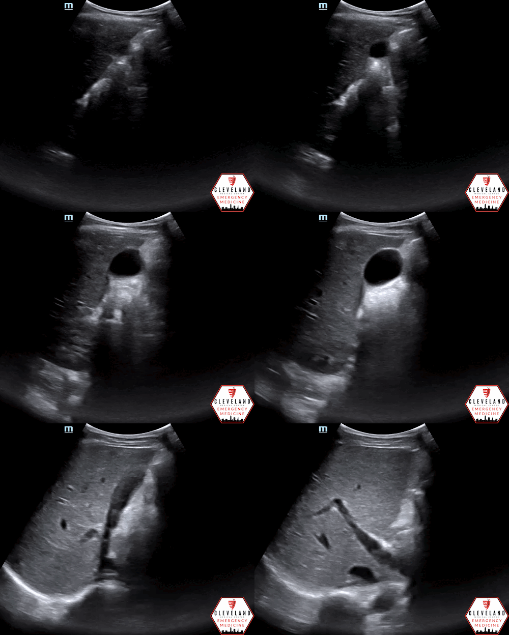
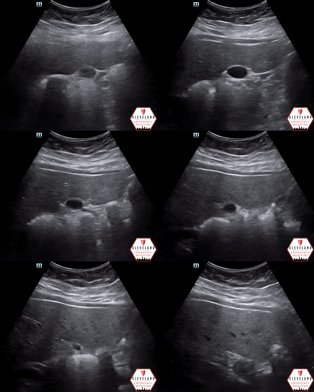
A biliary point-of-care ultrasound was performed to evaluate for gallstones or signs of cholecystitis.
POCUS findings: The gallbladder was well-visualized and there was no evidence of gallstones, increased wall thickness, pericholecystic fluid, common bile duct (CBD) dilation, or sonographic Murphy’s sign. Overall, grossly normal findings.
Case continued: Labs were obtained and unremarkable, further suggesting against biliary disease as well as pancreatitis. The patient received famotidine and Maalox and her pain improved, raising suspicion for GERD vs gastritis as possible etiologies for her pain. She was provided with reassurance and discharged home with instructions to follow up with her primary doctor.
While POCUS did not reveal any abnormal findings, it quickly ruled out cholelithiasis/cholecystitis, which helped reassure the patient/provider and expedite time to this patient’s disposition.
Cholecystitis: Brief Background
EPIDEMIOLOGY
Acute cholecystitis is the most common complication of cholelithiasis and typically develops in patients with a history of symptomatic gallstones.
Develops in 6 to 11 percent of patients with symptomatic gallstones over a median follow-up of 7 to 11 years [1]
The incidence of acute cholecystitis is approximately 6,300 per 100,000 in individuals under 50 years age and 20,900 per 100,000 in individuals over 50 years age worldwide [2]
Cholecystitis can be further subclassified into:
Acute — accounts for 85-90% of cases
Chronic — 10-15% of cases
CLINICAL SIGNS
Variable. May be ill-appearing, febrile, tachycardic, and lying still
Often have RUQ pain/tenderness. Positive Murphy’s Sign (sensitivity 97%, specificity 48%) [4]
Lab abnormalities may include:
Leukocytosis +/- bandemia
Transaminitis (usually mild)
Elevated lipase (if pancreatitis is present)
Hyperbilirubinemia and elevated alkaline phosphatase levels are less common, especially in the absence of CBD obstruction [5-6]
***But, lab values are nonspecific and may be normal
POCUS Assessment for Acute Cholecystitis
Focused Clinical Questions
Are gallstones present?
Are there signs of cholecystitis? (see below for specific findings)
Is the CBD dilated?
TECHNIQUE AND FINDINGS [3,7,8]
Curvilinear probe (alternatively: phased array)
Patient supine, knees flexed (if possible)
Left lateral decubitus or upright positioning can help optimize views
Find the gallbladder
Anterior approach: Place probe in epigastrium in longitudinal orientation. The goal is to identify 3 fluid filled structures. First, identify the aorta. As you slide to the right, the IVC will come into view (compressible and runs flush with the liver). As you continue to slide your probe, the third fluid filled structure will be the gallbladder.
Lateral approach: place probe in mid-axillary line (RUQ FAST view). Locate the hepatorenal interface and slowly slide/fan the probe anteriorly until able to visualize the gallbladder
X minus 7 approach: uses intercostal space as a window to view the gallbladder. The probe is placed ~7cm lateral to the xiphoid process, and the probe oriented to maximize the window between rib spaces.
If the gallbladder is not identified, continue to move the probe laterally while fanning through the liver parenchyma
Once you’ve identified the gallbladder, find its longest axis. This will typically require some degree of probe rotation as the gallbladder often sits at an oblique angle relative to the long axis of the patient’s body
As with most organs, it is important to visualize the gallbladder in two planes (rotate probe 90 degrees to obtain). Fan all the way through the gallbladder in both planes.
***The key area to visualize adequately is the neck of the gallbladder as this is where stones tend to get stuck and cause obstruction.
Measure the anterior gallbladder wall (short axis is preferred to avoid an oblique measurement which is more likely to happen in a long axis view)
<3mm is normal
Identify portal triad = portal vein, CBD and hepatic artery
In a longitudinal view of the gallbladder, the portal triad is often viewed adjacent to the gallbladder, known as “Mickey Mouse” sign
Identify the CBD, which lies anterior to the portal vein, has brighter walls, and no flow with Color Doppler. Can visualize in its long or short axis.
Measure inner wall to inner wall.
Source: http://lluultrasound.org/home/ebook/abdo
Some Troubleshooting Tips
If bowel gas is getting in the way: place patient in a left lateral decubitus or upright position — this helps move the bowel away from the gallbladder
If rib shadows are a problem: have the patient perform a deep inspiratory hold — this will move the diaphragm (and abdominal contents including the gallbladder) caudally, away from the ribs.
If you’re still having difficulty identifying the gallbladder, try a different approach or position
Evaluate for the following:
Sonographic Murphy Sign
Considered positive if the point of maximal tenderness in the right upper quadrant is identified while the gallbladder is visualized on ultrasound
Gallstones
Appear as hyperechoic structures in the gallbladder lumen with posterior shadowing
Gallstones that are not impacted may cause biliary colic that resolves spontaneously
If impacted in the neck of the gallbladder or CBD they can cause bile stasis and subsequent cholecystitis
Repositioning the patient can be helpful in determining whether stones are impacted or mobile
Anterior Wall Thickening
Abnormal wall thickness is defined as > 3mm
Measured at the thickest aspect of the wall
POCUS findings must be applied in conjunction with clinical reasoning, as there are a number of reasons why the gallbladder wall could be thickened; generally, this finding is associated with inflammation
Pericholecystic fluid
Appears as hypoechoic free fluid surrounding the gallbladder
Most commonly seen in the space between the gallbladder and liver
CBD dilation
While the literature is not totally consistent in defining normal CBD width, the general consensus states that a CBD >6 millimeters is considered distended
Some literature supports addition of 1mm per decade of life over the age of 60 [9]
Post-cholecystectomy the CBD can measure up to 1 centimeter normally [3]
Common pitfalls:
Failing to successfully recognize the structures of interest
Not repositioning the patient or attempting different techniques when the gallbladder is difficult to visualize
Mistaking duodenum/bowel for the gallbladder. Unlike a stone, bowel will have no shadowing or dirty shadowing and there may be peristalsis [10].
May have diffuse wall thickening in states of renal or liver failure, HIV, etc. [11]
WES (wall echo shadow) sign — gallbladder is contracted around multiple stones or single large stone, distorting normal appearance [12]
What Does the Evidence Show?
Emergency physician-performed biliary ultrasound has similar diagnostic accuracy as radiology-performed ultrasound in detecting cholelithiasis/cholecystitis [13]
CBD dilation is suggestive of biliary obstruction but utility is controversial [9]
Biliary POCUS has demonstrated reduced ED length-of-stay compared to radiology-performed ultrasonography [14]
Take Home Points
Biliary POCUS can quickly assess for cholelithiasis/cholecystitis with accuracy comparable to radiology-performed studies but is not as comprehensive
Learn the different approaches & troubleshooting tips as there is a lot of variable amongst patients
Assess gallbladder in its long AND short axis, identify anatomic structures (gallbladder & portal triad), & fan all the way through, focusing in particular on the gallbladder neck. Look for the above pathology & always measure the anterior GB wall thickness & CBD diameter.
AUTHORED BY: DR. NICK DIMEO, PGY1
FACULTY CO-AUTHOR/EDITOR: LAUREN MCCAFFERTY, MD
REFERENCES
Friedman GD. Natural history of asymptomatic and symptomatic gallstones. Am J Surg. 1993;165(4):399-404.
Kimura Y, et al. Definitions, pathophysiology, and epidemiology of acute cholangitis and cholecystitis: Tokyo Guidelines. J Hepatobiliary Pancreat Surg. 2007; 14 (1): 15–26.
Estrella A (2019). Biliary Ultrasound. Core EM. Ed. Vermeulen M. Retrieved Oct 2021 from: https://coreem.net/core/biliary-ultrasound/
Singer AJ, McCracken G, Henry MC, Thode HC Jr, Cabahug CJ. Correlation among clinical, laboratory, and hepatobiliary scanning findings in patients with suspected acute cholecystitis. . Ann Emerg Med. 1996;28(3):267.
Kurzweil SM, Shapiro MJ, Andrus CH, Wittgen CM, Herrmann VM, Kaminski DL. Hyperbilirubinemia without common bile duct abnormalities and hyperamylasemia without pancreatitis in patients with gallbladder disease. Arch Surg. 1994;129(8):829.
Moscati RM. Cholelithiasis, cholecystitis, pancreatitis. Emerg Med Clin North Am. 1996;14(4):719-37.
Avila, J. (2020). Gallbladder. Core Ultrasound. Retrieved Oct 2021 from:: https://www.coreultrasound.com/gallbladder/
UCSF ED Liver and Gallbladder/Biliary Ultrasound Protocol. UCSF Emergency Medicine. Retrieved Oct 2021 from: https://edus.ucsf.edu/sites/edus.ucsf.edu/files/wysiwyg/UCSF%20ED%20US%20Protocol%20Biliary_Final.pdf
Lahham S, Becker BA, Gari A, Bunch S, Alvarado M, Anderson CL, et al. Utility of common bile duct measurement in ED point of care ultrasound: A prospective study. Am J Emerg Med. 2018;36(6):962-966.
Smith B (2014). USOTW #8 answer. Core Ultrasound. Retrieved Oct 2021 from: https://www.coreultrasound.com/uotw-8-answer/
Runner GJ, Corwin MT, Siewert B, Eisenberg RL. Gallbladder wall thickening. Am J Roentgenol. 2014; 202(1): 202:W1–W12.
Miller AH, Pepe PE, Brockman CR, Delaney KA. ED ultrasound in hepatobiliary disease. J Emerg Med. 2006; 30(1):69–74.
Summers SM, Scruggs W, Menchine MD, et al. A prospective evaluation of emergency department bedside ultrasonography for the detection of acute cholecystitis. Ann Emerg Med. 2010;56(2):114-122.
Blaivas M, Harwood RA, Lambert MJ. Decreasing length of stay with emergency ultrasound examination of the gallbladder. Acad Emerg Med. 1999;6:1020-1023.


