POCUS: Small Bowel Obstruction
The Case
63yo F with history of CVA earlier this year resulting in PEG tube dependence presented for 12 hrs of nausea, bilious emesis, diffuse abdominal pain, and inability to tolerate PO, preceded by two days of reported obstipation. She was non-toxic but uncomfortable-appearing. Had a distended and diffusely tender abdomen though not peritonitic. Labs and CT were ordered.
In the meantime, POCUS was performed to assess for SBO and revealed the following:


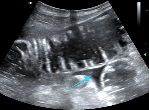

POCUS findings: fluid-filled, dilated loops of bowel with to-and-fro movement of contents, plicae circulares ("keyboard sign"), wall thickening, all suggestive of SBO. ***Note that normal bowel isn't nearly this well-visualized.
In the 3rd image, you see free fluid (arrow) between loops of bowel, which is associated with higher grade obstruction and poorer prognosis. This is referred to as the “tanga sign” as the fluid often assumes a triangular shape similar to that of a bikini bottom (#funfact)
Case continued: Diagnosis was confirmed a few hours later with CT, which showed distal obstruction. The patient was decompressed and admitted, ultimately managed conservatively though still in the hospital 2 weeks later.
Evaluating for SBO with POCUS
Technique
Curvilinear probe
Place probe in transverse orientation (marker to pt right) in RLQ or LLQ and scan up and down moving toward the opposite side ("mowing the lawn" approach)
Perform sequential, graded compression (gentle downward pressure every few cm) to assess for bowel compressibility and findings of SBO
US findings suggestive of SBO:
Fluid-filled bowel, diameter >2.5 cm
"To-and-fro" movement -- decreased or absent peristalsis
Well-defined plicae circulares extending perpendicularly from bowel wall (absent in ileum) - "keyboard sign"
Bowel wall edema
Distally collapsed bowel suggests a transition point
Free fluid between loops of bowel - "tanga sign"
*Free fluid is associated with higher grade obstruction and poorer prognosis
Some pitfalls:
Don't confuse with large bowel --- which has haustra (echogenic curvilinear arcs of colonic wall giving it a segmental appearance)
Air (such as from air-fluid levels or perforation) may result in A-lines and preclude a good view of bowel. If this is the case, consider a more lateral or inferior approach since air tends to layer in the least dependent areas (anteriorly if supine or superiorly if upright)
Often difficult to differentiate SBO from ileus (visualization of a transition point favors SBO)
Body habitus, as always
Why does this matter?
High sensitivity with generally lower but variable specificity compared to CT. Overall much better than KUB
When performed by providers with more ultrasound training, the diagnostic accuracy increased
Bottom Line
While POCUS doesn't replace CT, it can give you a diagnosis within minutes (literally) rather than hours and can help guide/expedite management.
POST BY: DR. LAUREN MCCAFFERTY
References
https://www.coreultrasound.com/small-bowel-obstruction/
https://www.coreultrasound.com/uotw-20-answer/
Ogata M. General Surgery Applications. In OJ Ma, JR Mateer, RF Reardon, SA Joing (eds), Ma and Mateer’s Emergency Ultrasound (3rd ed). New York, NY: McGraw-Hill Education. pp 273-317.
Abu-Zidan, F.M., Cevik, A.A. Diagnostic point-of-care ultrasound (POCUS) for gastrointestinal pathology: state of the art from basics to advanced. World J Emerg Surg. 2018; 13, 47.
Hefny AF, Corr P, Abu-zidan FM. The role of ultrasound in the management of intestinal obstruction. J Emerg Trauma Shock. 2012;5(1):84-6.
Taylor MR, Lalani N. Adult small bowel obstruction. Acad Emerg Med. 2013;20(6):528-44.
Grassi R, Romano S, D’amario F, et al. The relevance of free fluid between intestinal loops detected by sonography in the clinical assessment of small bowel obstruction in adults. Eur J Radiol. 2004;50(1):5-14
Pourmand A, Dimbil U, Drake A, Shokoohi H. The Accuracy of Point-of-Care Ultrasound in Detecting Small Bowel Obstruction in Emergency Department. Emerg Med Int. 2018;2018:3684081.
Becker BA, Lahham S, Gonzales MA, Nomura JT, Bui MK, Truong TA, et al. A Prospective, Multicenter Evaluation of Point-of-care Ultrasound for Small-bowel Obstruction in the Emergency Department. Acad Emerg Med. 2019 Aug;26(8):921-930.
Jang T. B., Schindler D., Kaji A. H. Bedside ultrasonography for the detection of small bowel obstruction in the emergency department. Emerg Med J. 2011;28(8):676–678