An Expanding Lung Mass & Assessment for Consolidations on Lung Ultrasound
The Case
A 65-year-old female with a past medical history including stage IV lung cancer for which she is receiving palliative radiation therapy, heart failure, and COPD presented to the emergency department for acute on chronic shortness of breath over the past few days. She also reported months of intermittent chest pain and cough but otherwise denied any new symptoms.
On exam, she was noted to be uncomfortable-appearing, tachypneic, tachycardic with low normal BP. She had a slight increase in her oxygen requirement compared to baseline of 2L NC. She had diminished breath sounds bilaterally and tenderness to palpation along the right sternal border.
She was given duonebs and steroids for possible COPD exacerbation. A workup was initiated while POCUS of her lungs and heart were performed.

Area of consolidation is seen in the right upper lung.


Normal right upper lung adjacent to the lung mass. The rest of the right lung and all the visualized aspects of the left lung appear similar to this.

Normal right lung base (similar to the RUQ view of the FAST exam. No effusion or consolidation are seen.
POCUS Findings: A focal consolidation is seen in the right upper chest (near site of known lung mass), resulting in disruption of the pleural line. It has a tissue-like appearance with focal b-lines but no dynamic air bronchograms (making pneumonia less likely). The lung immediately adjacent to this is normal-appearing, further suggesting a localized lesion. The remaining visualized areas of the right lung as well as the left lung were grossly unremarkable. Cardiac ultrasound showed reduced EF consistent with her baseline and was also otherwise unremarkable.
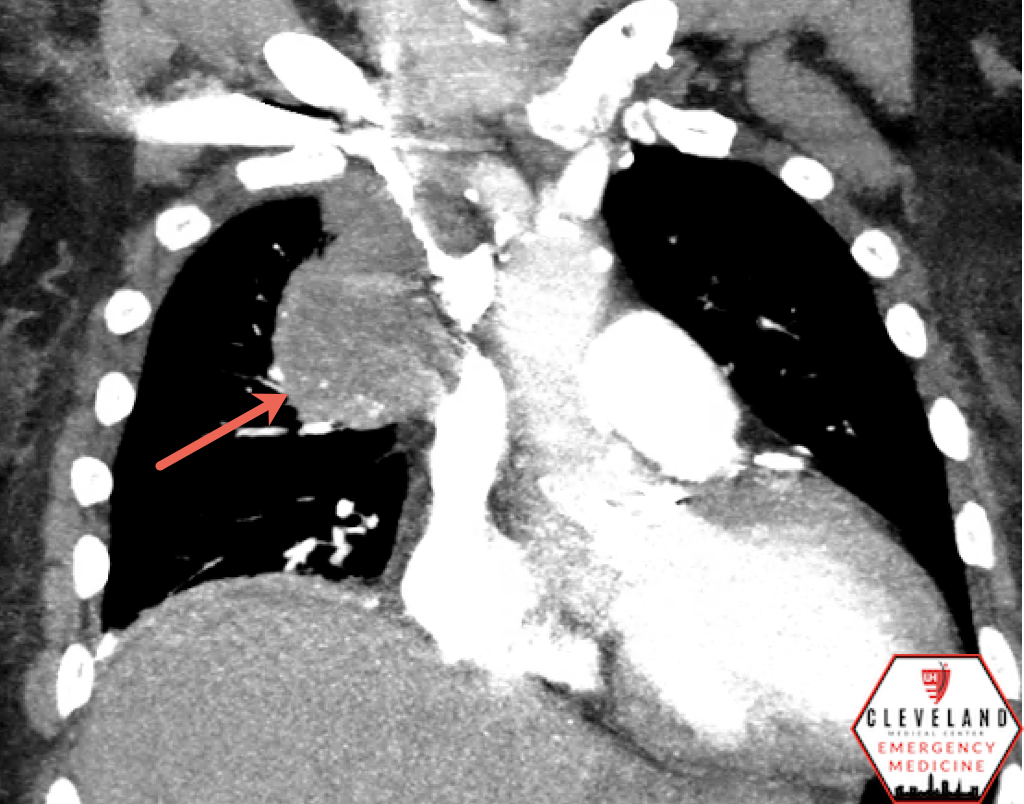
Case Continued: A CT scan of her chest was obtained and showed interval progression of right upper lung mass; there was no evidence for a PE or other acute process. Her respiratory distress improved and she remained stable while in the ED. She was admitted to the medicine service for her increased oxygen requirement and symptomatic management. The patient and family ultimately decided to transition to palliative care.
POCUS Assessment for Lung Consolidations
*Lung ultrasound is a broad topic. The focus here will be on consolidations.
Indications
Any patient with shortness of breath, pleuritic chest pain, focal findings on lung auscultation, or other signs/symptoms concerning for lung pathology.
*Because patients may have very diverse presentations given the variety of pathology that can result in a lung consolidation, clinician gestalt and clinical context is important.
Technique & Findings
Probe Selection
Curvilinear is best but the phased-array works as well. The linear probe should be reserved for assessment of the pleural line (not as good for assessing deeper structures such as larger consolidation).
Patient Positioning
Patients are most commonly assessed in a supine position, which allows for evaluation of the anterior and lateral lung fields. If the patient is able to sit upright, this will also allow for assessment of the posterior aspects of the lung [1-3].
Areas of Assessment/Probe Placement
In general, an adequate lung ultrasound exam assesses a minimum of four lung fields – the right upper, right lower, left upper, and left lower lung fields [4]. However, the more areas of the lung assessed (including the posterior aspects of the lung), the more comprehensive the study. This helps when differentiating diffuse vs focal pathology and is especially important when assessing for consolidations; a more limited exam may miss smaller, more focal consolidations or other findings. Additionally, assessment in both sagittal and transverse orientations improves diagnostic yield for consolidations [5].
The diagrams below are a helpful way to organize the fields in your mind. If a specific zone is suspected based on physical exam or prior imaging, a more detailed assessment of that area is recommended.

Platz et. al. 2019 [4]

http://www.emdocs.net/lung-ultrasound-in-covid-19/
For the more simplified approach:
Assess the upper lung fields by placing the probe over the anterior chest near the midclavicular line over the 2-3rd intercostal space with the probe marker pointing toward the patient’s head.
Assess the lower lung fields by placing the probe in the mid-posterior axillary line (similar to the RUQ/LUQ views for the FAST exam). Visualize the basilar aspect of the lung where it meets the diaphragm. Best view to assess for pleural effusion.
As mentioned above, a more comprehensive exam looking at more lung fields, particularly the posterior zones, improves the diagnostic accuracy when assessing for consolidation.
*Tip: Because lung ultrasound relies primarily on artifacts for both normal and abnormal findings, keep the probe perpendicular to the chest wall to allow for appropriate generation of artifacts [1-2].
Ultrasound Findings
Normal Findings [1-3]
Lung sliding is the shimmering movement of the hyperechoic pleural line resulting from the visceral pleural moving against the parietal pleura.


A-lines are a type of reverberation artifact generated from sound waves bouncing back and forth between the highly reflective pleural line and the transducer, resulting in repetitive horizontal lines at regularly spaced intervals. They simply represent the presence of air. Because air scatters sound waves, a normal air-filled lung will prevent visualization of any other structures deep to the pleural line.


Consolidations [1-3, 6-8]
As alveoli accumulate fluid, this area of the lung is able to transmit the sound waves, these actually show up as distinct areas of abnormal tissue within the lung, often adjacent to normal areas of lung.
Depending on the disease process, findings of consolidation may include:
“Hepatization” or tissue-like appearance — consolidated lung takes on an echogenic appearance that mimics a solid organ such as the liver
Air bronchograms — hyperechoic areas within consolidated lung that represent air trapped in smaller airways
Dynamic — movement with respiration. Considered pathognomonic for pneumonia.
Static — lack of movement. Can be seen in other types of consolidation such as atelectasis
Shred sign — represents irregular border of a consolidation
B-lines (focal) and/or pleural effusion — may be seen focally near the consolidation

Hepatization with static air bronchograms with associated focal b-lines & pleural effusion

DYNAMIC air bronchograms

Consolidation with irregular borders (Shred sign) with disruption of the pleural line [1]
Differential includes:
Pneumonia (lobar): Hepatization, air bronchograms, and focal b-lines are often present. Dynamic air bronchograms are essentially diagnostic but not very sensitive [6-7]. There may be an associated pleural effusion in the lower lung field on the same side as the consolidation.
In contrast, viral pneumonia tends to lack a large consolidation but instead has a thickened/irregular pleural line and subpleural consolidation(s), along with focal b-lines; pleural effusions are rare [9]
Atelectasis — may appear similar to pneumonia but will lack dynamic air bronchograms; only static air bronchograms may be present [7]. If it’s compressive atelectasis (such as from a pleural effusion), the borders tend to be “cleaner”/more regular-appearing.
Malignancy — may also have a tissue-like appearance, static air bronchograms, disruption of the pleural line, increased internal vascularity. May have invasion into the chest wall itself and involve adjacent structures [10]
Infarct — usually well-defined wedge-shaped subpleural consolidation, may have surrounding effusion [11]
Compressive atelectasis from the surrounding pleural effusion (“jelly fish sign”)

Infarct from a PE - Triangular shaped hypoechoic region disrupting the pleural line [1]
Less commonly seen
Tuberculosis — Findings may include apical consolidations, subpleural consolidations, or multiple consolidations (in disseminated disease) rather than a single large mass. Diagnostic accuracy of lung ultrasound for TB findings is not well-established. [12]
Asthma: Incredibly diverse range of findings. Completely normal lung ultrasound findings are common, as small airway inflammation does not always pass the threshold of edema to cause b-lines or other focal findings. If airway edema is significant, there may be diffuse b-lines. However, if a focal area has significant inflammation, a segment of lung may become walled off from the upper airway. Though less common, a focal consolidation may be present on US in up to 30% of cases [13]
*Note: Other types of rare lesions may present with very similar ultrasound findings and can be difficult to differentiate. Clinical judgement should always be used in conjunction with ultrasound findings.
**B-lines and pleural effusions are generally less specific and can be seen in a number of other disease processes. Not discussed in detail here, but their distribution can help differentiate. For pulmonary edema, b-lines are typically diffusely distributed and predominantly in the more dependent areas of the lung [1-3].
Advantages & Supporting Evidence for Lung US for Pneumonia
Lung ultrasound has been shown to have significantly greater sensitivity and specificity compared to chest x-ray [14]. For consolidations detectable by CT, ultrasound has demonstrated specificity of 83% and specificity of 96% [15]. Similarly, a systemic review and meta-analysis found ultrasound to have a pooled sensitivity of 85% and specificity of 93% compared to either CXR or CT [16]. Because point-of-care ultrasound is portable and can be performed at the bedside, it is a quicker, repeatable, and more cost-effective tool to assess for pathology.
Limitations
Like all POCUS applications, lung ultrasound is operator dependent. Additionally, bony landmarks such as the scapula can obscure certain areas of the lung fields making a truly complete scan of all lung tissue difficult. While US is relatively sensitive for abnormalities closer to the surface of the pleura, artifact and machine limitations can make visualizing deeper consolidations difficult [1-3].
Take Home Points
When suspecting pathologies with focal lung findings like pneumonia or malignancy, consider a more thorough assessment, evaluating as many lung fields as possible.
Dynamic air bronchograms, when present, are highly specific for pneumonia. Hepatization, shred sign, focal b-lines, associated pleural effusion are other common findings
Most pathologies associated with consolidations may be difficult to differentiate with ultrasound findings alone so clinical context should be taken into account.
Consider POCUS to assess for pneumonia or other consolidations. While it may not replace the need for other imaging, it may give you an answer more quickly which can expedite management. Similarly, if you have a chest x-ray and it’s non diagnostic, POCUS may be able to differentiate.
AUTHORED BY: DR. DYLAN SEXTON (PGY1)
FACULTY CO-AUTHOR/EDITOR: LAUREN MCCAFFERTY, MD
References:
1. Silva FR, Mills LD. Pulmonary. In OJ Ma, JR Mateer, RF Reardon, SA Joing (eds), Ma and Mateer’s Emergency Ultrasound (3rd ed). McGraw-Hill Education. pp 169-190.
2. Marini TJ, et. al. Lung Ultrasound: The Essentials. Radiol Cardiothorac Imaging. 2021 ;3(2):e200564.
3. Mojoli F., Bouhemad B., Mongodi S., Lichtenstein D. Lung Ultrasound for Critically Ill Patients. Am J Respir Crit Care Med. 2019;199(6):701-714.
4. Platz E, et al. Lung ultrasound in acute heart failure: Prevalence of pulmonary congestion and short- and long-term outcomes. JACC Heart Fail. 2019; 7(10): 849-858.
5. Milliner BH, Tsung JW. Lung consolidation locations for optimal lung ultrasound scanning in diagnosing pediatric pneumonia. J Ultrasound Med. 2017;36(11):2325-2328.
6. Gillman LM, Panebianco N, Alkadi A, Blaivas M, Kirkpatrick AW. The Dynamic Sonographic Air Bronchogram: A Simple and Immediate Bedside Diagnosis of Alveolar Consolidation in Severe Respiratory Failure. J Trauma. 2011; 70(3):760.
7. Lichtenstein D, Mezière G, Seitz J. The dynamic air bronchogram. A lung ultrasound sign of alveolar consolidation ruling out atelectasis. Chest. 2009;135(6):1421-1425.
8. Volpicelli G., Caramello V., Cardinale L, Mussa A, Bar F, Frascisco MF. Detection of sonographic B-lines in patients with normal lung or radiographic alveolar consolidation. Med Sci Monit. 2008 Mar;14(3):CR122-8.
9. Peng QY, Wang XT, Zhang LN, Critical C, Ultrasound C, Group S. Findings of lung ultrasonography of novel corona virus pneumonia during the 2019 – 2020 epidemic. Intensive Care Med. 2020;(87):6-7.
10. Hafez MR, Sobh ESM, Elsawy SB, Abo-Eikheir OA. The usefulness of thoracic ultrasonography in diagnosis and staging of bronchogenic carcinoma. Ultrasound. 2017; 25(4): 200–212.
11. Reissig A, Heyne JP, Kroegel C. Sonography of lung and pleura in pulmonary embolism: Sonomorphologic characterization and comparison with spiral CT scanning. Chest. 2001; 120(6):1977–1983.
12. Montuori M., Casella F., et. al. Lung Ultrasonography in pulmonary tuberculosis: A pilot study on diagnostic accuracy in a high-risk population. Eur J Int Med. 2019; 66; (29-34)
13. Dankoff, S., Li P., et. al. Point of care lung ultrasound of children with acute asthma exacerbations in the pediatric ED. American Journal of Emergency Medicine 2017 Apr;35(4):615-622.
14. Corellaro F, Colombo S, Coen D, Duca PG. Lung ultrasound is an accurate diagnostic tool for the diagnosis of pneumonia in the emergency department. Emerg Med J. 2012;29(1):19-23.
15. Nazerian P. et. al. Accuracy of lung ultrasound for diagnosis of consolidations when compared to chest computed tomography. Am J Emerg Med. 2015; 33(5) 620-625.
16. Alzahrani SA, Al Salamah MA, Al-Madani WH, Eibarbary MA. Systematic review and meta-analysis for the use of ultrasound versus radiology in diagnosing of pneumonia, Crit Ultrasound J. 2017; 9(1):6.