Intern Ultrasound of the Month: Superficial Thrombophlebitis
The Case
Middle aged male with active tonsillar cancer being treated with chemoradiation presented to the ED for 2 days of progressive pain, redness, and swelling in his left forearm. Symptoms started slightly below his antecubital fossa and gradually spread distally to his wrist.. Had a peripheral IV in his left AC 5 days prior to onset of symptoms though which he received IV chemo. No systemic symptoms. ROS otherwise negative. Of note, he is on long-term warfarin for prior DVTs but currently being held for recent peg tube placement.
He arrived to the ED hemodynamically stable, afebrile, well-appearing. Exam notable for well-demarcated erythema and warmth over ventral aspect of left forearm extending from ~2cm below AC to his wrist. Tender to palpation over the affected area, slightly worse centrally. No open wounds, fluctuance, palpable masses. Neurovascularly intact. No joint involvement.
Point-of-care ultrasound findings
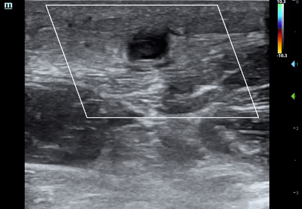
Llnear probe in short axis over the mid forearm revealed a superficial, well-circumscribed, hypoechoic structure with intraluminal echogenicity that was not compressible.
Also viewed in long axis (not shown), it extended horizontally across the entire screen, suggesting a vessel rather than abscess/mass.

Color flow applied and no flow within the structure was noted
The structure was traced proximally, a few centimeters distal to AC with similar findings.

Moving proximally in long axis, you can see that this structure converges with another, supporting the notion that this is a vessel.

Continuing more proximally (in short axis), just above the AC, the vessel lumen became fully anechoic and flow visualized. Though not shown here, it was also fully compressible, thus confirming that this is a vein with thrombosis distally. Pulsatile artery seen on the right of the screen in far field. Deep vein adjacent to it.

Diagnosis: Superficial Thrombophlebitis
—Ultrasound Findings, Pearls & Pitfalls—
Direct visualization of thrombus
Lack of compressibility
Lack of color flow
Don’t confuse with abscess (you don’t want to cut into this!!!)
Trace proximally & use color doppler to confirm it’s a vessel
“Squish sign” (swirling/movement of contents with compression) suggests abscess
Differentiate vein vs artery (look proximal to thrombus)
Compressibility? — favors vein but depends on volume status
Wall thickness? — thicker wall favors arteries but can be difficult to assess in distal vessels
Pulsatility? — favors arteries but also depends on volume status (***use light continuous compression to better assess, 5minsono_artvsvein)
Apply color doppler
Signs of infection (thrombophlebitis tends to be inflammatory)
Cobblestoning —> cellulitis
Dirty shadows —> emphysema (suggesting necrotizing infection)
Superficial Thrombophlebitis
What is it?: pain/ inflammation + thrombus within the superficial venous system
Pain & inflammation of vein but no thrombus = phlebitis
Part of broader category of superficial venous thrombosis

https://commons.wikimedia.org/wiki/File:Veins_of_the_forearm_and_hand.jpg
Risk Factors: female, older age, vessel cannulation, hypercoagulable states, hyper/hypotonic or irritant infusions (including chemotherapeutics)
Signs/symptoms: erythema, warmth, tenderness of affected area. May have palpable cord. May develop systemic sx if suppurative
***Mimics cellulitis, milder necrotizing infection, DVT, abscess
Diagnosis: primarily clinical, but ultrasound helps assess clot burden/location & evaluate for mimickers
Prognosis: usually self-limited but consider severity and comorbidities
Consider re-evaluated in 7-10 days to assess for progression
Treatment: elevate extremity, warm compresses, NSAIDs
Antibiotics? not routinely indicated but consider patient comorbidities, signs of infection, overall clinical status
Risk for venous thromboembolism (DVT, PE)?
Upper extremities: very low risk but poorly studied Cochrane Review 2015
Lower extremities: slightly increased risk, primarily if larger clot (>5cm), non-varicose veins, or hypercoagulable state/high risk Cochrane Review 2018
These patients may benefit from low dose anticoagulation x 45 days but evidence is controversial (must weight benefit with bleed risk)
Clot within 3cm of saphenofemoral junction in lower extremities should be treated like DVT
Back to the case…
Patient diagnosed with superficial thrombophlebitis based on soft tissue involvement with likely occluded cephalic vein based on location/distribution. He was provided with warm compresses and pain control. Given his immunocompromised status, basic labs obtained and unremarkable. Based on the progression of his symptoms, immunocompromised status, and recommendation by his oncologist, he was admitted for pain control and observation. While inpatient, the primary team ordered a DVT ultrasound study which was negative. His home warfarin, which had been held for recent peg tube placement, was restarted (given his prior DVTs). Patient did well, symptoms started to improve, and he was discharged two days later.
Conclusions/Why Does POCUS Matter?
Color Doppler and compression are your friends!
Rule out abscess & avoid I&D!
POCUS confirms clinical diagnosis & helps guide management
Differentiate from abscess, cellulitis, necrotizing infection, etc.
May avoid more advanced diagnostic imaging (time, radiation, cost, etc)
Avoid unnecessary antibiotics
Can expedite time to diagnosis & disposition
Just another example of POCUS for the win! :)
POST BY: DR. JOSIAH SMITH, PGY1
FACULTY EDITING BY: DR. LAUREN MCCAFFERTY
References
Beyer-WestendorfJ. Controversies in venous thromboembolism: to treat or not to treat superficial vein thrombosis. Hematology Am SocHematolEducProgram. 2017;2017(1):223–230.M DN, Im W, Middeldorp S. Treatment for superficial thrombophlebitis of the leg. Cochrane Database of Systematic Reviews 2018; (2).
Nisio DM, Aws R. Cochrane Database of Systematic Reviews Treatment for superficial infusion thrombophlebitis of the upper extremity (Review). 2015;(11).
Zehnder, James MD. Catheter-related upper extremity venous thrombosis. UpToDate. Waltham, MA: UpToDate Inc. https://www.uptodate.com
http://blog.5minsono.com/artvsvein/
http://5minsono.com/svt/